. 2012 Mar;30(3):452-60.
doi: 10.1002/stem.1013.

RNA-binding protein L1TD1 interacts with LIN28 via RNA and is required for human embryonic stem cell self-renewal and cancer cell proliferation

Affiliations

RNA-binding protein L1TD1 interacts with LIN28 via RNA and is required for human embryonic stem cell self-renewal and cancer cell proliferation

Elisa Närvä et al. Stem Cells. 2012 Mar.

Abstract

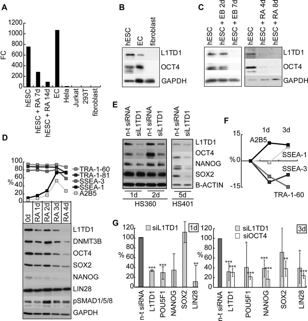
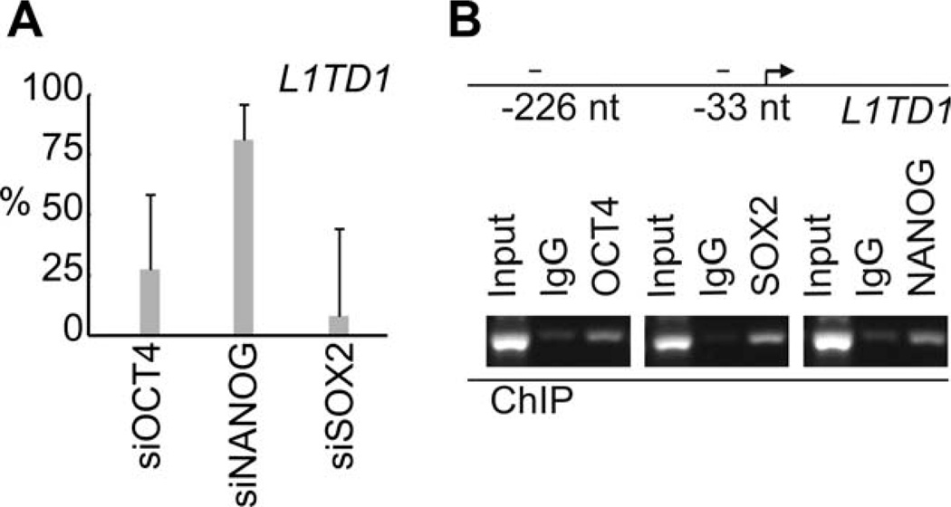
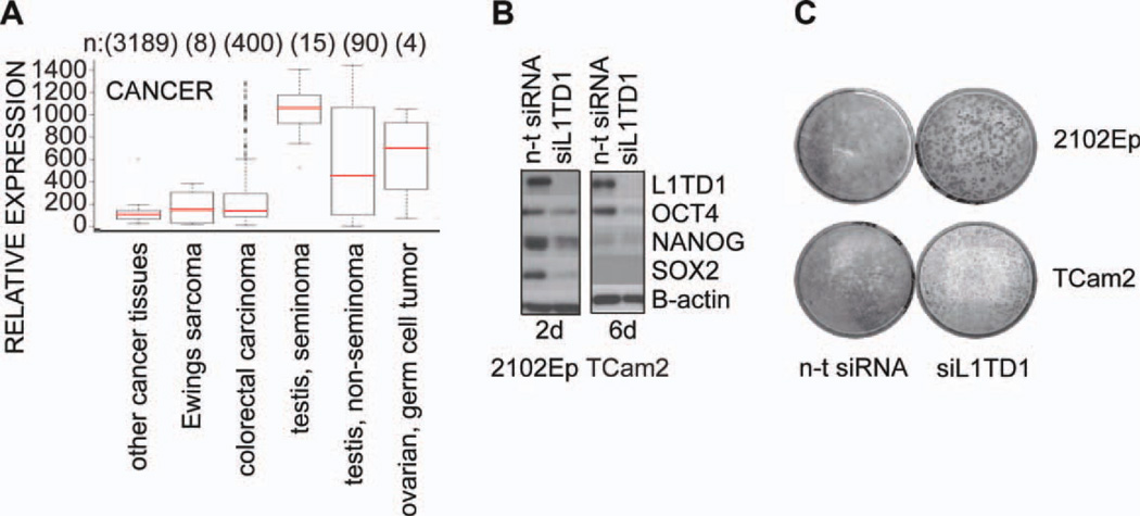
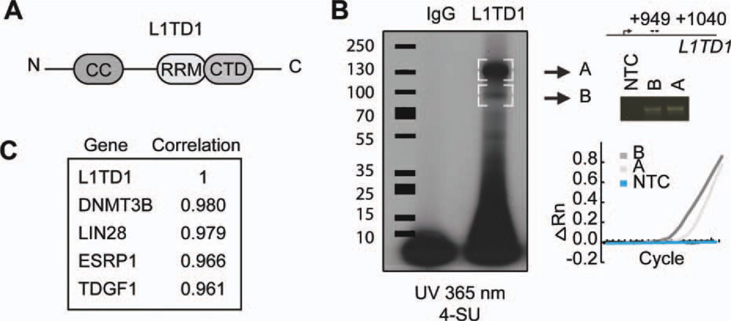
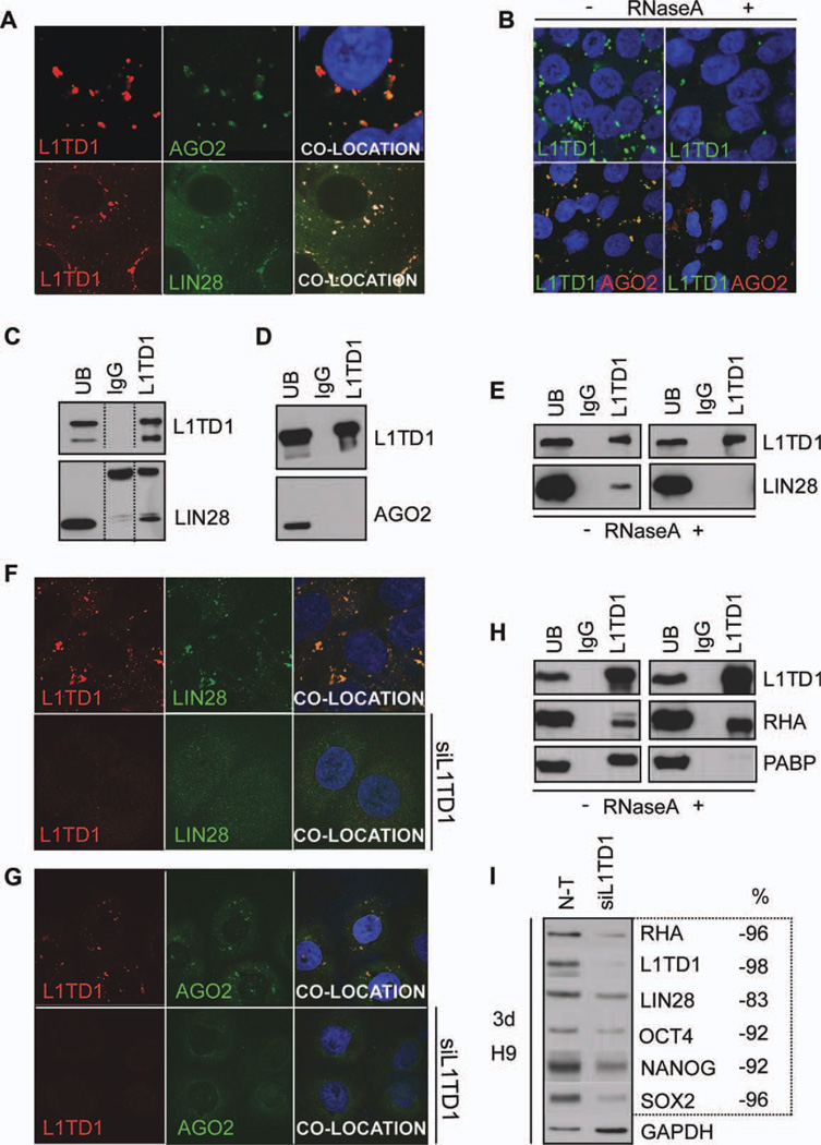
Human embryonic stem cells (hESC) have a unique capacity to self-renew and differentiate into all the cell types found in human body. Although the transcriptional regulators of pluripotency are well studied, the role of cytoplasmic regulators is still poorly characterized. Here, we report a new stem cell-specific RNA-binding protein L1TD1 (ECAT11, FLJ10884) required for hESC self-renewal and cancer cell proliferation. Depletion of L1TD1 results in immediate downregulation of OCT4 and NANOG. Furthermore, we demonstrate that OCT4, SOX2, and NANOG all bind to the promoter of L1TD1. Moreover, L1TD1 is highly expressed in seminomas, and depletion of L1TD1 in these cancer cells influences self-renewal and proliferation. We show that L1TD1 colocalizes and interacts with LIN28 via RNA and directly with RNA helicase A (RHA). LIN28 has been reported to regulate translation of OCT4 in complex with RHA. Thus, we hypothesize that L1TD1 is part of the L1TD1-RHA-LIN28 complex that could influence levels of OCT4. Our results strongly suggest that L1TD1 has an important role in the regulation of stemness.


Figures

Figure 1
Figure 1
L1TD1 is highly expressed in hESCs and required for self-renewal. (A): Real-time PCR analysis of L1TD1 expression in hESCs (HS401), RA-induced differentiation (HS401), EC cells (NTERA2), Hela, Jurkat, 293T, and human fibroblasts. (B): Western blot analysis of L1TD1 and OCT4 expression in hESCs (H9), ECs (2102Ep), and human fibroblasts. (C): Western blot analysis of L1TD1 and OCT4 expression in EB and RA-induced differentiation of hESC (H9 and HS401, respectively). (D): Flow cytometry and Western blot analysis of selected pluripotency and differentiation markers in RA-induced differentiation (hESC line H9). Average data from two replicate cultures. (E): Western blot analysis of the OCT4, SOX-2, and NANOG 1, 2 (oligo 5), and 5 (oligo 2) days after siRNA knockdown of L1TD1 (hESC lines HS360 and HS401). (F): Flow cytometry analysis of pluripotency (SSEA-3, TRA-1-60) and differentiation (A2B5 and SSEA-1) markers 1 and 3 days after knockdown of L1TD1 in relation to the n-t control (hESC line H9) (siRNA oligo 5). Average data from two replicate cultures. (G): Real-time PCR analysis of L1TD1 and pluripotency markers: POU5F1 (OCT4), NANOG, SOX2, and LIN28 1 and 3 days after knockdown of L1TD1. Average data of hESC lines: H9 and HS360 for day 1 (siRNA oligo 1 and 5), H9 and HS237 for day 3 (siRNA oligos 2 and 5). p values: *, ≤ 0.05; **, ≤ 0.01; ***, ≤ 0.001. Abbreviations: EB, embryoid body; EC, embryonal carcinoma; FC, fold change; hESC, human embryonic stem cell; n-t, nontargeting; PCR, polymerase chain reaction; RA, retinoic acid; siRNA, small interfering RNA.
Figure 2
Figure 2
Expression of L1TD1 is regulated by core stem cell factors. (A): Expression of L1TD1 measured with real-time PCR after knockdown (KD) of OCT4 (KD-88%), NANOG (KD-75%), and SOX2 (KD-33%). Average data from days 1, 3, and 4 (human embryonic stem cells [hESC] line H9). (B): ChIP-PCR analysis of OCT4, SOX-2, and NANOG occupancy at L1TD1 locus (hESC line HS360). Abbreviations: ChIP, chromatin immunoprecipitation; PCR, polymerase chain reaction.
Figure 3
Figure 3
L1TD1 is necessary for self-renewal of embryonal carcinoma (EC) cells. (A): Relative expression of L1TD1 in cancer tissues in in silico transcriptomics database [18]. (B): Western blot analysis of expression of indicated proteins and their response to L1TD1 depletion in 2102Ep and TCam2 cells (siRNA oligo 2). (C): Colony forming assay performed with nonseminoma EC cell line (2102Ep) and seminoma cell line (TCam2) after silencing with n-t siRNA or siL1TD1 (siRNA oligo 2). Cells were stained with crystal violet 12 days post-transfection. Abbreviations: n-t, nontargeting; siRNA, small interfering RNA.
Figure 4
Figure 4
L1TD1 is an RNA-binding protein. (A): Structure of L1TD1 protein. N-terminal CC domain, an RRM, and a CTD. (B): SDS-PAGE autoradiography of 4-SU-labeled RNA after L1TD1 immunoprecipitation. Embryonic carcinoma cell line 2102Ep. Real-time PCR and gel analysis of L1TD1 bound L1TD1 messenger RNA extracted from gel bands A and B. (ΔRn = Rn − baseline), Rn = The ratio of the fluorescence emission intensity of the reporter dye to the fluorescence emission intensity of the passive reference dye. (C): The genes with the highest correlation of gene expression profiles with L1TD1 in Stem Cell Matrix data [9]. Abbreviations: CC, coiled coil; CTD, C-terminal domain; NTC, non-template control; RRM, RNA recognition motif; SDS-PAGE, sodium dodecyl sulfate polyacrylamide gel electrophoresis; SU, thiouridine.
Figure 5
Figure 5
L1TD1 localizes in P-bodies and interacts with LIN28 and RHA. (A): L1TD1 (red) colocalization with AGO2 (green) or LIN28 (green) (cell line HS360). (B): Effect of RNase A treatment on the localization of L1TD1 and AGO2 (cell line HS360). (C): Coimmunoprecipitation analysis of L1TD1 and LIN28 (hESC line HS360). (D): Coimmunoprecipitation analysis of L1TD1 and AGO2 (NTERA2 cell line). (E): Effect of RNase A treatment on the coimmunoprecipitation of L1TD1 and LIN28 (2102Ep cell line). (F): Detection of L1TD1 (red) and LIN28 (green) in the presence or absence of siL1TD1 treatment (siRNA oligo 5) (hESC line H9). (G): Detection of L1TD1 (red) and AGO2 (green) in the presence or absence of siL1TD1 treatment (siRNA oligo 2) (TCam2 cell line). (H): Coimmunoprecipitation analysis of L1TD1 with RHA and PABP in the presence and absence of RNase A (2102Ep cell line). (I): Western blot analysis of the expression of indicated proteins and their response to L1TD1 depletion (siRNA oligo 5). Proportion (%) of the protein expression relative to that in the n-t siRNA. The values have been normalized with the housekeeping protein expression (GAPDH). Abbreviations: n-t, nontargeting; PABP, poly(A)-binding protein; RHA, RNA helicase A; UB, unbound fraction.

References

    1. Boyer LA, Lee TI, Cole MF, et al. Core transcriptional regulatory circuitry in human embryonic stem cells. Cell. 2005;122:947–956. - PMC - PubMed
    1. Kuersten S, Goodwin EB. The power of the 30 UTR: Translational control and development. Nat Rev Genet. 2003;4:626–637. - PubMed
    1. Lee MH, Schedl T. RNA-binding proteins. WormBook. 2006:1–13. - PMC - PubMed
    1. Mitsui K, Tokuzawa Y, Itoh H, et al. The homeoprotein Nanog is required for maintenance of pluripotency in mouse epiblast and ES cells. Cell. 2003;113:631–642. - PubMed
    1. Wong RC, Ibrahim A, Fong H, et al. L1TD1 is a marker for undifferentiated human embryonic stem cells. PLoS One. 2011;6:e19355. - PMC - PubMed

Publication types

MeSH terms