

Regional differences in the evolution of lung disease in children with cystic fibrosis
- PMID: 22162514
- PMCID: PMC3310260
- DOI: 10.1002/ppul.21604
Regional differences in the evolution of lung disease in children with cystic fibrosis
Abstract
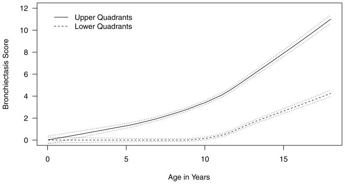
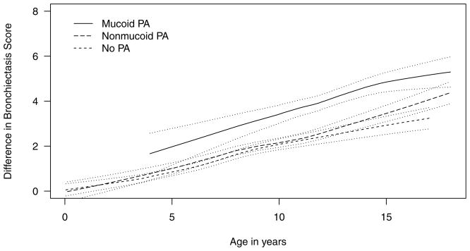
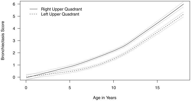
Progression of lung disease is a major event in children with cystic fibrosis (CF), but regional differences in its evolution are unclear. We hypothesized that regional differences occur beginning in early childhood. We examined this issue by evaluating 132 patients followed in the Wisconsin Neonatal Screening Project between 1985 and 2010. We scored chest X-rays obtained every 1-2 years with the Wisconsin chest X-ray system, in which we divided the lungs into quadrants, and gave special attention to ratings for bronchiectasis (BX) and nodular/branching opacities. We compared the upper and lower quadrant scores, and upper right and left quadrant scores, as patients aged using a multivariable generalized estimation equation (GEE) model. We did a confirmatory analysis for a subset of 81 patients with chest computerized tomography (CT) images obtained in 2000 and scored using the Brody scoring system. The chest X-ray analysis shows that the upper quadrants have higher BX (P<0.001) and nodular/branching opacities (P<0.001) scores than the lower quadrants. CT analysis likewise reveals that the upper quadrants have more BX (P=0.02). Patients positive for mucoid PA showed significantly higher BX scores than patients with non-mucoid PA (P=0.001). Chest X-ray scoring also revealed that the upper right quadrant has more BX (P<0.001) than the upper left quadrant, and CT analysis was again confirmatory (P<0.001). We conclude that pediatric patients with CF develop more severe lung disease in the upper lobes than the lower lobes in association with mucoid PA infections and also have more severe lung disease on the right side than on the left side in the upper quadrants. A variety of potential explanations such as aspiration episodes may be clinically relevant and provide insights regarding therapies.
Copyright © 2011 Wiley Periodicals, Inc.
Figures
Similar articles
-
[New method of scoring lung changes using computed tomography in patients with cystic fibrosis].Med Wieku Rozwoj. 2012 Oct-Dec;16(4):290-302. Med Wieku Rozwoj. 2012. PMID: 23378408 Polish.
-
Chest computed tomography scores of severity are associated with future lung disease progression in children with cystic fibrosis.Am J Respir Crit Care Med. 2011 Oct 1;184(7):816-21. doi: 10.1164/rccm.201105-0816OC. Am J Respir Crit Care Med. 2011. PMID: 21737586 Free PMC article.
-
Prospective longitudinal association between repeated multiple breath washout measurements and computed tomography scores in children with cystic fibrosis.J Cyst Fibros. 2021 Jul;20(4):632-640. doi: 10.1016/j.jcf.2020.09.010. Epub 2020 Oct 4. J Cyst Fibros. 2021. PMID: 33028501
-
Scoring of chest CT in children with cystic fibrosis: state of the art.Pediatr Radiol. 2014 Dec;44(12):1496-506. doi: 10.1007/s00247-013-2867-y. Epub 2014 Aug 28. Pediatr Radiol. 2014. PMID: 25164326 Review.
-
What did we learn from two decades of chest computed tomography in cystic fibrosis?Pediatr Radiol. 2014 Dec;44(12):1490-5. doi: 10.1007/s00247-014-2964-6. Epub 2014 Aug 28. Pediatr Radiol. 2014. PMID: 25164327 Review.
Cited by
-
Cystic Fibrosis and Pseudomonas aeruginosa: the Host-Microbe Interface.Clin Microbiol Rev. 2019 May 29;32(3):e00138-18. doi: 10.1128/CMR.00138-18. Print 2019 Jun 19. Clin Microbiol Rev. 2019. PMID: 31142499 Free PMC article. Review.
-
Cheating by type 3 secretion system-negative Pseudomonas aeruginosa during pulmonary infection.Proc Natl Acad Sci U S A. 2014 May 27;111(21):7801-6. doi: 10.1073/pnas.1400782111. Epub 2014 May 12. Proc Natl Acad Sci U S A. 2014. PMID: 24821799 Free PMC article.
-
Quantifying Spatial Distribution of Ventilation Defects in Multiple Pulmonary Diseases With Hyperpolarized 129Xenon MRI.J Magn Reson Imaging. 2025 Apr;61(4):1860-1873. doi: 10.1002/jmri.29627. Epub 2024 Oct 22. J Magn Reson Imaging. 2025. PMID: 39434582 Free PMC article.
-
Preliminary comparison of normalized T1 and non-contrast perfusion MRI assessments of regional lung disease in cystic fibrosis patients.J Cyst Fibros. 2017 Mar;16(2):283-290. doi: 10.1016/j.jcf.2015.11.009. Epub 2015 Dec 22. J Cyst Fibros. 2017. PMID: 26719281 Free PMC article.
-
A Flow Cytometric Method for Isolating Cystic Fibrosis Airway Macrophages from Expectorated Sputum.Am J Respir Cell Mol Biol. 2019 Jul;61(1):42-50. doi: 10.1165/rcmb.2018-0236MA. Am J Respir Cell Mol Biol. 2019. PMID: 30742539 Free PMC article.
References
-
- Ramsey B. Management of pulmonary disease in patients with cystic fibrosis. N Engl J Med. 1996;335(3):179–188. - PubMed
-
- Maffessanti M, Candusso M, Brizzi F, Piovesana F. Cystic fibrosis in children: HRCT findings and distribution of disease. J Thorac Imaging. 1996;11(1):27–38. - PubMed
-
- Santis G, Hodson ME, Strickland B. High resolution computed tomography in adult cystic fibrosis patients with mild lung disease. Clin Radiol. 1991;44(1):20–22. - PubMed
-
- Davis SD, Fordham LA, Brody AS, Noah TL, Retsch-Bogart GZ, Qaqish BF, Yankaskas BC, Johnson RC, Leigh MW. Computed tomography reflects lower airway inflammation and tracks changes in early cystic fibrosis. Am J Respir Crit Care Med. 2007;175(9):943–950. - PubMed
-
- Cleveland R, Neish A, Zurakowski D, Nichols D, Wohl M, Colin A. Cystic fibrosis: predictors of accelerated decline and distribution of disease in 230 patients. AJR Am J Roentgenol. 1998;171(5):1311–1315. - PubMed
Publication types
MeSH terms
Grants and funding
LinkOut - more resources
Full Text Sources
Medical
