. 2011:3:25.
doi: 10.3410/B3-25. Epub 2011 Dec 1.

The puzzling origin of the autophagosomal membrane

The puzzling origin of the autophagosomal membrane

Muriel Mari et al. F1000 Biol Rep. 2011.

Abstract

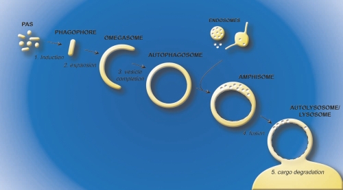
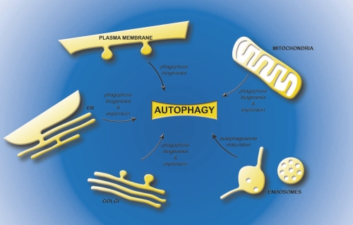
Autophagy is one of the newest and fastest emerging research areas in biomedical life sciences. Autophagosomes, large double-membrane vesicles enclosing cytoplasmic components targeted for degradation, are the hallmark of this catabolic pathway. The origin of the lipid bilayers composing these transport carriers has been the central enigma of the field since the discovery of autophagy. A series of recent studies has implicated several cellular organelles as the possible source of the autophagosomal membranes, if anything further clouding our view. In this compendium, we will discuss these apparently contradictory results and briefly emphasize the relevance of determining the lipid source used for autophagy for future translational research, for example in drug discovery programs.


Figures

Figure 1.
Figure 1.. Model for autophagosome biogenesis and cargo degradation
The process of autophagy can be divided in five steps. The induction (1) is elicited by the formation of the phagophore (or isolation membrane), which then expands (2) around the material targeted for degradation. The omegasome probably represents a phagophore expansion intermediate. The closure of the growing phagophore (3) leads to the formation of a complete autophagosome. The autophagosome then fuses with endosomal structures to become an amphisome, an organelle specific to high eukaryotic cells, where the sequestered material starts to be degraded. Subsequently, the amphisome fuses (4) with lysosomes (vacuoles in plants and yeast) to generate autolysosomes, in the interior of which resident hydrolases break down the internal membrane of autophagosomes (dashed lines) and the cargo into basic metabolites (5). These metabolites are finally transported in the cytoplasm where they are reused as either a source of energy or building blocks for new proteins and lipids. Abbreviations: PAS, phagophore assembly site.
Figure 2.
Figure 2.. A working definition of the membrane carriers mediating autophagy
A defined order for the Atg (autophagy-related) protein recruitment to the phagophore assembly site has been established in yeast [18] and recent results in mammalian cells suggest that this hierarchy is conserved [17]. Based on the available experimental data, it is possible to use a few proteins to begin to molecularly define the various autophagosomal intermediates. The distribution of the following marker proteins is shown: Atg14L is a subunit of the phosphatidylinositol-3-kinase complex involved in autophagy [60]. FIP200 associates with ULK1/2, Atg13, Atg101 into the ULK1/2 complex [61]. DFCP-1 (double FYVE-containing protein 1) could represent the marker protein that allows us to distinguish omegasomes from phagophores [30]. Atg12, Atg5 and Atg16 form a complex, e.g. Atg12-5-16, which predominantly localizes to the phagophore, but Atg16L may have an even earlier role in autophagy [41]. Lipidated LC3, e.g. LC3-II, and Lamp-1 (lysosomal-associated membrane protein 1) are well-characterized marker proteins of the late compartments of autophagy. Note that LC3-II is present in the interior of the amphisome/autophagosome and sensitive to lysosomal hydrolyases while Lamp-1 is an integral membrane protein and resistant to these hydrolyases. Abbreviations: Atg, autophagy-related; DFCP1, double FYVE-containing protein 1; LAMP1, lysosomal-associated membrane protein 1.
Figure 3.
Figure 3.. Possible sources of the lipids bilayers for the different intermediates of autophagy
The plasma membrane, the endoplasmic reticulum, the mitochondria and the Golgi have been proposed to contribute to phagophore biogenesis. In addition, the endoplasmic reticulum, Golgi and mitochondria, could also provide the membranes necessary for the phagophore expansion, while endosomal compartments are necessary for the maturation of autophagosomes into amphisomes. Abbreviations: ER, endoplasmic reticulum.

References

    1. Maiuri MC, Zalckvar E, Kimchi A, Kroemer G. Self-eating and self-killing: crosstalk between autophagy and apoptosis. Nat Rev Mol Cell Biol. 2007;8:741–52. doi: 10.1038/nrm2239. - DOI - PubMed
    1. Kroemer G, Marino G, Levine B. Autophagy and the integrated stress response. Mol Cell. 2010;40:280–93. doi: 10.1016/j.molcel.2010.09.023. - DOI - PMC - PubMed
    1. Mizushima N, Levine B. Autophagy in mammalian development and differentiation. Nat Cell Biol. 2010;12:823–30. doi: 10.1038/ncb0910-823. - DOI - PMC - PubMed
    1. Levine B, Deretic V. Unveiling the roles of autophagy in innate and adaptive immunity. Nat Rev Immunol. 2007;7:767–77. doi: 10.1038/nri2161. - DOI - PMC - PubMed
    1. Deretic V, Levine B. Autophagy, immunity, and microbial adaptations. Cell Host Microbe. 2009;5:527–49. doi: 10.1016/j.chom.2009.05.016. - DOI - PMC - PubMed