

Emphysematous cystitis: radiological diagnosis of complicated urinary tract infection
- PMID: 22162733
- PMCID: PMC3028265
- DOI: 10.1136/bcr.05.2009.1832
Emphysematous cystitis: radiological diagnosis of complicated urinary tract infection
Abstract
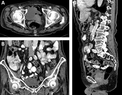
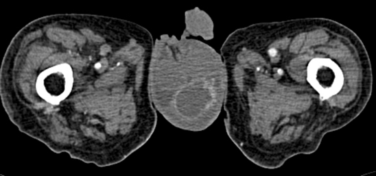
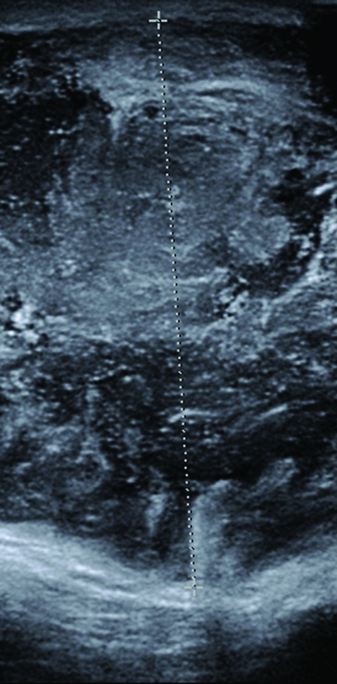
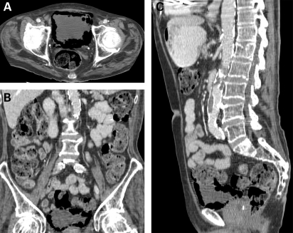
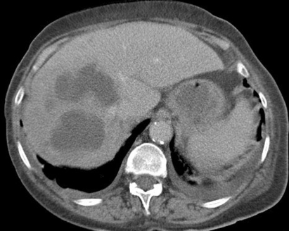
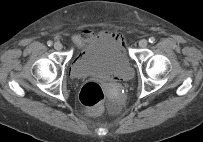
Emphysematous cystitis is an uncommon condition characterised by the presence of gas in the bladder. It is an infection caused by gas forming organisms, usually in elderly women with a background of diabetes mellitus. The presentation is variable, however with increasing use of imaging more cases are being diagnosed in asymptomatic patients. Routine cross-sectional imaging is not advocated for specific diagnosis but its role in accurate assessment of the severity of the condition cannot be overlooked. As the mode and duration of follow-up in incidentally detected cases has not been addressed in the literature, follow-up should be tailored individually depending upon the severity and response to treatment. We describe two such incidentally detected cases of emphysematous cystitis in elderly diabetic patients and present a review of the literature. The triad of treatment is adequate control of diabetes, antibiotics and bladder drainage. One patient died in the hospital, while the other underwent a flexible cystoscopy 6 weeks later which was normal.
Figures
References
-
- Thomas AA, Lane BR, Thomas AZ, et al. Emphysematous cystitis: a review of 135 cases. BJU Int 2007; 100: 17–20 - PubMed
-
- Mokabberi R, Ravakhah K. Emphysematous urinary tract infections: diagnosis, treatment and survival (case review series). Am J Med Sci 2007; 333: 111–16 - PubMed
-
- Grupper M, Kravtsov A, Potasman I. Emphysematous cystitis: illustrative case report and review of the literature. Medicine 2007; 86: 47–53 - PubMed
-
- Stapleton A. Urinary tract infections in patients with diabetes. Am J Med 2002; 113(Suppl 1A): 80S–84S - PubMed
-
- Tseng CC, Wu JJ, Wang MC, et al. Host and bacterial virulence factors predisposing to emphysematous pyelonephritis. Am J Kidney Dis 2005; 46: 432–9 - PubMed
LinkOut - more resources
Full Text Sources
Research Materials