

Gene transfer using micellar nanovectors inhibits choroidal neovascularization in vivo
- PMID: 22162776
- PMCID: PMC3230610
- DOI: 10.1371/journal.pone.0028560
Gene transfer using micellar nanovectors inhibits choroidal neovascularization in vivo
Abstract
Purpose: Age-related macular degeneration caused by choroidal neovascularization (CNV) remains difficult to be treated despite the recent advent of several treatment options. In this study, we investigated the in vivo angiogenic control by intravenous injection of polyion complex (PIC) micelle encapsulating plasmid DNA (pDNA) using a mice CNV model.
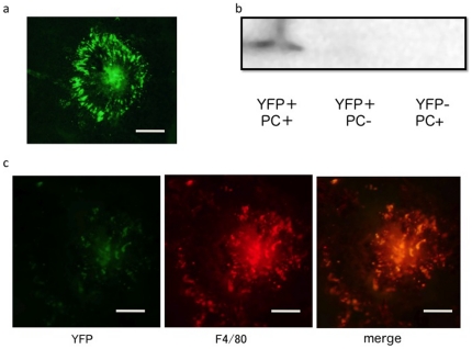
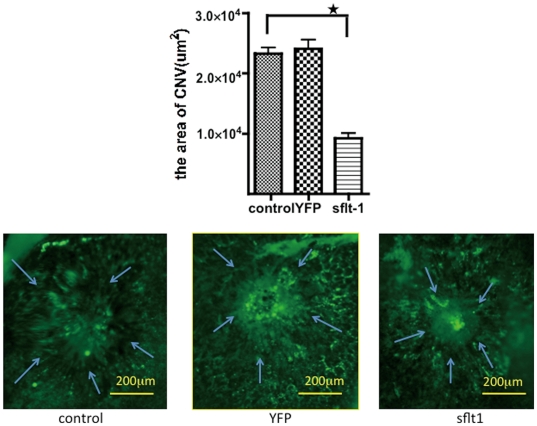
Methods: The transfection efficiency of the PIC micelle was investigated using the laser-induced CNV in eight-week-old male C57 BJ/6 mice. Firstly, each mouse received intravenous injection of micelle encapsulating pDNA of Yellow Fluorescent Protein (pYFP) on days 1,3 and 5. The expression of YFP was analyzed using fluorescein microscopy and western blotting analysis. In the next experiments, each mouse received intravenous injection of micelle encapsulating pDNA of soluble Fms-like tyrosine kinase-1 (psFlt-1) 1,3 and 5 days after the induction of CNV and the CNV lesion was analyzed by choroidal flatmounts on day 7.
Results: Fluorescein microscopy and western blotting analysis revealed that the expression of YFP was confirmed in the CNV area after injection of the PIC micelle, but the expression was not detected neither in mice that received naked pDNA nor those without CNV. Furthermore, the CNV area in the mice that received intravenous injection of the psFlt-1-encapsulated PIC micelle was significantly reduced by 65% compared to that in control mice (p<0.01).
Conclusions: Transfection of sFlt-1 with the PIC micelle by intravenous injection to mice CNV models showed significant inhibition of CNV. The current results revealed the significant potential of nonviral gene therapy for regulation of CNV using the PIC micelle encapsulating pDNA.
Conflict of interest statement
Figures
References
-
- Ferris FL, Fine SL, Hyman L. Age-related macular degeneration and blindness due to neovascular maculopathy. Arch Ophthalmol. 1984;102:1640–1642. - PubMed
-
- Wormald R, Evans J, Smeeth L. Cochrane Database Syst Rev; 2000. Photodynamic therapy for neovascular age-related macular degeneration.CD002030 - PubMed
-
- Rosenfeld PJ, Brown DM, Heier JS, Boyer DS, Kaiser PK, et al. Ranibizumab for neovascular age-related macular degeneration. N Engl J Med. 2006;355:1419–1431. - PubMed
-
- Gragoudas ES, Adamis AP, Cunningham ET, Jr, Feinsod M, Guyer DR. Pegaptanib for neovascular age-related macular degeneration. N Engl J Med. 2004;351:2805–2816. - PubMed
Publication types
MeSH terms
Substances
LinkOut - more resources
Full Text Sources
Medical
Miscellaneous
