

Physeal growth arrest by excessive compression: histological, biochemical, and micro-CT observations in rabbits
- PMID: 22162794
- PMCID: PMC3232359
- DOI: 10.4055/cios.2011.3.4.309
Physeal growth arrest by excessive compression: histological, biochemical, and micro-CT observations in rabbits
Abstract
Background: Compressive force across the growth plate may cause retardation and even arrest of physeal growth. The purpose of this study was to investigate histologic changes, metabolic changes in terms of glycosaminoglycan (GAG) concentration, and contrast-enhanced micro-computed tomography (CEMCT) findings of physeal cartilage in a rabbit model of physeal damage caused by excessive compression.
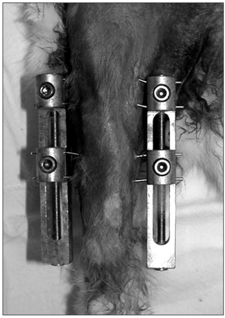
Methods: Compressive forces were applied via external fixators for two weeks to the growth plates of distal femurs and proximal tibiae of right hind-legs in 8-week-old rabbits. Left hind-legs remained intact and were used as controls. Forty-four bone specimens containing growth plates of distal femurs or proximal tibiae were harvested one week (n = 12) and four weeks (n = 32) after surgery, and examined for histologic findings (H&E staining) and GAGs quantification in physeal cartilage. After incubation in an ionic contrast material for 48 hours, specimens were scanned by CEMCT, and the pixel values of physeal cartilage were measured.
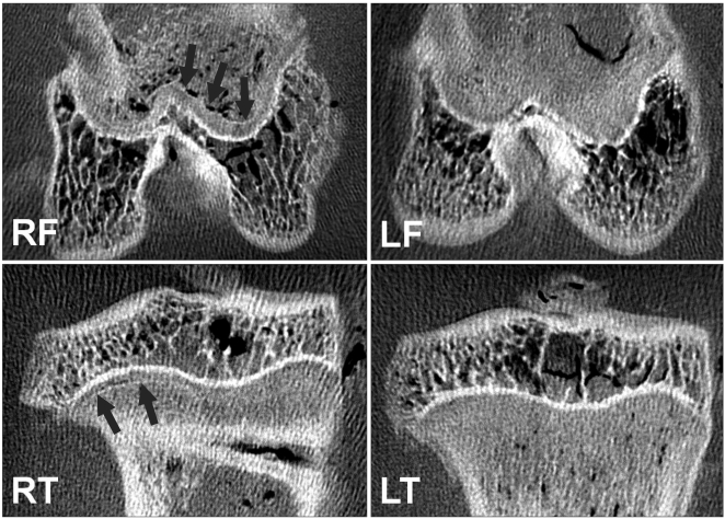
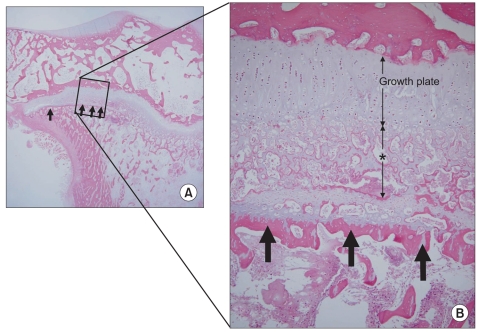
Results: CEMCT showed a thin, highly attenuated line parallel to the growth plate in compressed specimens harvested at four weeks after surgery, which was found to be transversely connected trabecular bone. In these specimens, GAG content in physeal cartilage was significantly lower, and CEMCT pixel values of physeal cartilage were significantly higher than in the specimens from the contralateral control side.
Conclusions: Excessive compressive force applied to growth plates produces altered histologic features and metabolic function in terms of decreased GAG content in physeal cartilage, changes that can be demonstrated by CEMCT.
Keywords: Contrast-enhanced micro-CT; Glycosaminoglycan; Growth arrest; Growth plate; Rabbit.
Conflict of interest statement
No potential conflict of interest relevant to this article was reported.
Figures
References
-
- Futami T, Foster BK, Morris LL, LeQuesne GW. Magnetic resonance imaging of growth plate injuries: the efficacy and indications for surgical procedures. Arch Orthop Trauma Surg. 2000;120(7-8):390–396. - PubMed
-
- Cockman MD, Blanton CA, Chmielewski PA, et al. Quantitative imaging of proteoglycan in cartilage using a gadolinium probe and microCT. Osteoarthritis Cartilage. 2006;14(3):210–214. - PubMed
-
- Piscaer TM, van Osch GJ, Verhaar JA, Weinans H. Imaging of experimental osteoarthritis in small animal models. Biorheology. 2008;45(3-4):355–364. - PubMed
-
- Farndale RW, Buttle DJ, Barrett AJ. Improved quantitation and discrimination of sulphated glycosaminoglycans by use of dimethylmethylene blue. Biochim Biophys Acta. 1986;883(2):173–177. - PubMed
Publication types
MeSH terms
LinkOut - more resources
Full Text Sources
