. 2012 May;46(5):592-8.
doi: 10.1165/rcmb.2011-0116OC. Epub 2011 Dec 8.

Long-term exposure of chemokine CXCL10 causes bronchiolitis-like inflammation

Affiliations

Long-term exposure of chemokine CXCL10 causes bronchiolitis-like inflammation

Dianhua Jiang et al. Am J Respir Cell Mol Biol. 2012 May.

Abstract

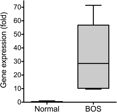
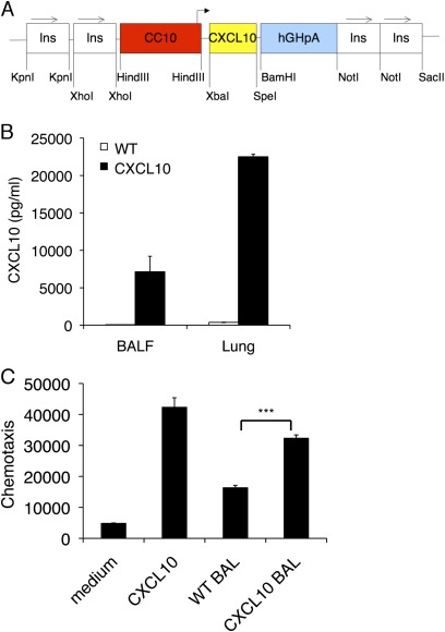
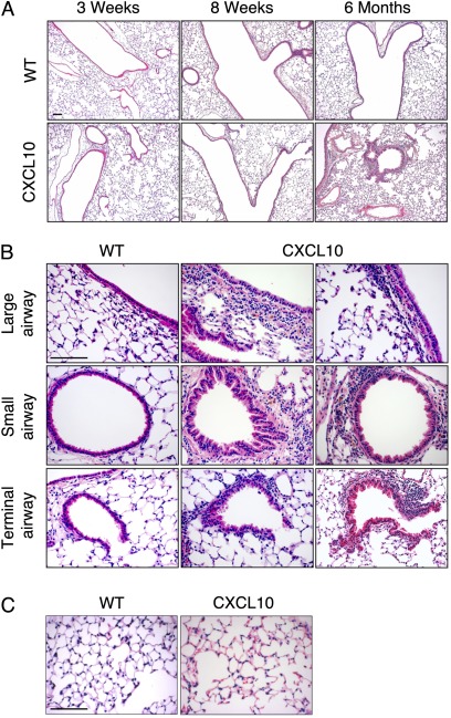
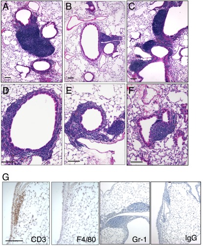
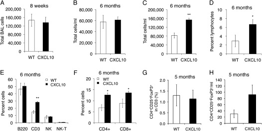
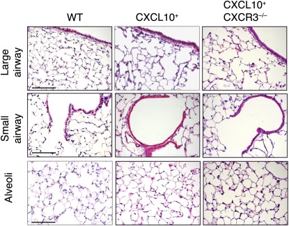
Chemokines and chemokine receptors have been implicated in the pathogenesis of bronchiolitis. CXCR3 ligands (CXCL10, CXCL9, and CXCL11) were elevated in patients with bronchiolitis obliterans syndrome (BOS) and chronic allorejection. Studies also suggested that blockage of CXCR3 or its ligands changed the outcome of T-cell recruitment and airway obliteration. We wanted to determine the role of the chemokine CXCL10 in the pathogenesis of bronchiolitis and BOS. In this study, we found that CXCL10 mRNA levels were significantly increased in patients with BOS. We generated transgenic mice expressing a mouse CXCL10 cDNA under control of the rat CC10 promoter. Six-month-old CC10-CXCL10 transgenic mice developed bronchiolitis characterized by airway epithelial hyperplasia and developed peribronchiolar and perivascular lymphocyte infiltration. The airway hyperplasia and T-cell inflammation were dependent on the presence of CXCR3. Therefore, long-term exposure of the chemokine CXCL10 in the lung causes bronchiolitis-like inflammation in mice.


Figures

Figure 1.
Figure 1.
Increased CXCL10 mRNA in the lungs of patients with bronchiolitis obliterans syndrome (BOS). Total RNA was isolated from the lung tissues of patients with BOS and from control subjects. RT-PCR was used to determine CXCL10 mRNA levels in the lung tissues. Housekeeping gene GusB mRNA levels were used a control (n = 3; P < 0.05).
Figure 2.
Figure 2.
Overexpression of CXCL10 in the lung. (A) Transgenic construct to show that the mouse CXCL10 cDNA was cloned downstream of the rat CC10 promoter and upstream of human growth hormone polyadenylation and intronic sequence. Two copies of the chicken β-globin insulator sequence were flanked on both ends of the transgene. (B) CC10–CXCL10 transgenic mice produced a large amount of CXCL10 protein into bronchoalveolar lavage (BAL) and lung tissue. CXCL10 levels in BAL and lung tissue in 8-week-old transgenic mice and their wild-type (WT) littermates were measure with ELISA (n = 6). (C) CXCL10 in BAL was bioactive. Jurkat cells were plated onto a Boyden chamber. BAL from wild-type or CXCL10 transgenic mice was added to the bottom chamber. Recombinant CXCL10 was used as a positive control. Cells that migrated to the bottom chamber were counted after 4 hours. (n = 3; ***P < 0.001).
Figure 3.
Figure 3.
Airway inflammation in CC10–CXCL10 transgenic mice. (A) Lung micrographs (H&E staining) of CC10–CXCL10 transgenic mice and wild-type (WT) littermate control mice at 3 weeks, 8 weeks, or 6 months. Airway inflammation was seen in CC10–CXCL10 transgenic mice at 6 months of age. Scale bar, 100 μm. (B) Lung histology (H&E) of CC10–CXCL10 transgenic mice and WT littermate control mice at 6 months. Upper panels, large airways; middle panels, small airways; lower panels, terminal airway. Scale bar, 100 μm. (C) Lung histology (H&E) of CC10–CXCL10 transgenic mice and WT littermate control mice at 6 months to demonstrate that there was no apparent alveolar inflammation. Scale bar, 100 μm.
Figure 4.
Figure 4.
Lymphocytic infiltration. (AF) Lung histology (H&E staining) of CC10–CXCL10 transgenic mice at 6 months of age displayed lymphocytic nodules around airways. (G) Lung sections of 6-month-old CC10–CXCL10 transgenic mice were stained with specific antibodies against T-cell marker CD3, macrophage marker F4/80, neutrophil marker Gr-1, or control IgG. Scale bar, 100 μm.
Figure 5.
Figure 5.
Lung inflammation. (A) Total BAL cells from CC10–CXCL10 transgenic mice and WT littermate control mice at 8 weeks of age (n = 4; P > 0.05). (B) Total BAL cells of CC10–CXCL10 transgenic mice and WT littermate control mice at 6 months of age (n = 4 or 5; P > 0.05). (C) Total mouse lung cells of CC10–CXCL10 transgenic mice and WT littermate control mice at 6 months of age (n = 4 or 5; P < 0.01). (D) Lymphocyte percentage of cell differential count of total mouse lung cells of CC10–CXCL10 transgenic mice and WT littermate control mice at 6 months of age (n = 4 or 5; P < 0.05). (E, F) Flow cytometry to determine lymphocytes in total mouse lung cells of CC10–CXCL10 transgenic mice and WT littermate control mice at 6 months of age (n = 4 or 5; *P < 0.05; **P < 0.01). (G) Flow cytometric analysis of regulatory T cells (CD4+CD25+FoxP3+) in BAL (n = 3 or 4; P > 0.05). (H) Total Treg cells in BAL (n = 3 or 4; P > 0.05).
Figure 6.
Figure 6.
Histology of CXCL10+CXCR3–/– mice. Lung micrographs (H&E) of CXCL10+CXCR3–/– mice, CXCR3–/– mice, and WT littermate control mice at 6 months of age. Normal airway structure (either large or small) was demonstrated in CXCL10+CXCR3–/– mice, and there was no leukocyte infiltration and no apparent alveolar inflammation in CXCL10+CXCR3–/– mice. Scale bar, 100 μm.

References

    1. AAP Diagnosis and management of bronchiolitis. Pediatrics 2006;118:1774–1793 - PubMed
    1. Popper HH. Bronchiolitis, an update. Virchows Arch 2000;437:471–481 - PubMed
    1. Cooper JD, Billingham M, Egan T, Hertz MI, Higenbottam T, Lynch J, Mauer J, Paradis I, Patterson GA, Smith C, et al. A working formulation for the standardization of nomenclature and for clinical staging of chronic dysfunction in lung allografts. International Society for Heart and Lung Transplantation. J Heart Lung Transplant 1993;12:713–716 - PubMed
    1. Snyder LD, Palmer SM. Immune mechanisms of lung allograft rejection. Semin Respir Crit Care Med 2006;27:534–543 - PubMed
    1. Mukae H, Kadota J, Kohno S, Kusano S, Morikawa T, Matsukura S, Hara K. Increase in activated CD8+ cells in bronchoalveolar lavage fluid in patients with diffuse panbronchiolitis. Am J Respir Crit Care Med 1995;152:613–618 - PubMed

Publication types