. 2011;6(12):e28457.
doi: 10.1371/journal.pone.0028457. Epub 2011 Dec 6.

IL-1α/IL-1R1 expression in chronic obstructive pulmonary disease and mechanistic relevance to smoke-induced neutrophilia in mice

Affiliations

IL-1α/IL-1R1 expression in chronic obstructive pulmonary disease and mechanistic relevance to smoke-induced neutrophilia in mice

Fernando M Botelho et al. PLoS One. 2011.

Abstract

Background: Cigarette smoking is the main risk factor for the development of chronic obstructive pulmonary disease (COPD), a major cause of morbidity and mortality worldwide. Despite this, the cellular and molecular mechanisms that contribute to COPD pathogenesis are still poorly understood.

Methodology and principal findings: The objective of this study was to assess IL-1 α and β expression in COPD patients and to investigate their respective roles in perpetuating cigarette smoke-induced inflammation. Functional studies were pursued in smoke-exposed mice using gene-deficient animals, as well as blocking antibodies for IL-1α and β. Here, we demonstrate an underappreciated role for IL-1α expression in COPD. While a strong correlation existed between IL-1α and β levels in patients during stable disease and periods of exacerbation, neutrophilic inflammation was shown to be IL-1α-dependent, and IL-1β- and caspase-1-independent in a murine model of cigarette smoke exposure. As IL-1α was predominantly expressed by hematopoietic cells in COPD patients and in mice exposed to cigarette smoke, studies pursued in bone marrow chimeric mice demonstrated that the crosstalk between IL-1α+ hematopoietic cells and the IL-1R1+ epithelial cells regulates smoke-induced inflammation. IL-1α/IL-1R1-dependent activation of the airway epithelium also led to exacerbated inflammatory responses in H1N1 influenza virus infected smoke-exposed mice, a previously reported model of COPD exacerbation.

Conclusions and significance: This study provides compelling evidence that IL-1α is central to the initiation of smoke-induced neutrophilic inflammation and suggests that IL-1α/IL-1R1 targeted therapies may be relevant for limiting inflammation and exacerbations in COPD.


Conflict of interest statement

Competing Interests: The authors have read the journal's policy and have the following conflicts: DF, SP, MS, RK, and AAH are full time employees of MedImmune, a wholly owned subsidiary of AstraZeneca. MLF is an employee of AstraZeneca. AJP is an employee of Pfizer. This does not alter the authors' adherence to all the PLoS ONE policies on sharing data and materials.

Figures

Figure 1
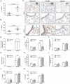
Figure 1. IL-1α and β are increased in the lung of chronic obstructive pulmonary disease patients.
Representative images of IL-1α (A) and β (B) expression in lung biopsies from GOLD I/II COPD patients. (C) Positive cells were enumerated from two biopsy samples obtained from each patient (n = 5 non-COPD and n = 9 COPD GOLD stage I/II patients). Statistical significance was determined using a Generalized Linear Mixed Effect model with negative binomial (adjusted for dispersion) to take into account multiple sampling of the same patient. Whiskers of box plots represent 1–99 percentile. Lung sections from the same biopsy samples were scored for IL-1α (D) and β (E) staining in the epithelium as follows: 0, no staining; 1, occasional staining; 2, marked focal staining; 3, marked diffuse staining. A stratified Wilcoxon Ranksum test was used to compare the frequencies of the staining categories (0, 1, 2, and 3) and represented graphically (size of block is proportional to frequency). Levels of IL-1α and β were measured in sputum samples obtained from patients at enrolment during stable disease (F), at onset of exacerbation (G), and days 7 (H) and 35 (I) post-exacerbation.
Figure 2
Figure 2. IL-1α and β expression in a smoke exposure model that induces IL-1R1-dependent and caspase-1-independent neutrophilia.
BALB/c mice were room air or cigarette smoke-exposed for 4 days. Total levels of IL-1α (A) and β (B) protein were measured by ELISA in lung homogenates (n = 5 mice per group). (C) Representative images showing expression of IL-1α and β in room air and smoke-exposed BALB/c mice. Insets represent macrophages from the interstitial space. Wild-type and either IL-1R1-deficient (C57BL/6 background) (n = 5 mice per group) (D–F) or caspase-1-deficient (G–I) mice (NOD/ShiLt background) (n = 3–6 mice per group) were room air or cigarette smoke-exposed for 4 days. Neutrophils (D and G) and mononuclear cells (F and I) were assessed in the broncho-alveolar lavage (BAL) of room air and smoke-exposed mice. Percentage of Gr-1hi cells were assessed by flow cytometric analysis of room air and smoke-exposed whole lung tissue from wild-type and either IL-1R1-deficient (E) (n = 5 mice per group) and caspase-1-deficient (H) (n = 4–6 mice per group) animals. Total levels of IL-1α (J) and β (K) protein were measured by ELISA from lung homogenates of room air and smoke-exposed wild-type and caspase-1-deficient mice (n = 4–6 mice per group). All data (B–K) are representative of two independent experiments.
Figure 3
Figure 3. Cigarette smoke-induced neutrophilia is IL-1α dependent and IL-1β-independent.
(A) 4 day cigarette smoke-exposed BALB/c mice were either left untreated (No Rx), or administered an isotype antibody (IgG isotype), or either an anti-IL-1α or anti-IL-1β blocking antibody. Neutrophil numbers were enumerated in the BAL (n = 4–5 mice per group from one of two independent experiments). (B) Wild-type and IL-1α-deficient mice were room air or cigarette smoke-exposed for 4 days. Data show BAL neutrophil numbers (n = 6–8 mice per group). CXCL-1 (C) and IL-1β (D) expression (left panels) were assessed in cigarette smoke-exposed and room air control BALB/c mice that were left untreated (No Rx), administered an isotype control antibody (IgG isotype), or either an anti-IL-1α or anti-IL-1β blocking antibody. Expression of cxcl-1 and IL-1β transcripts was assessed by fluidigm array and is presented relative to no treatment room air control animals (n = 5 mice per group from one of two independent experiments). Total protein levels (right panels) of CXCL-1 (C) and IL-1β (D) were measured by MSD (n = 10 mice per group from two independent experiments).
Figure 4
Figure 4. Neutrophilia induced by chronic cigarette smoke exposure is IL-1R1/IL-1α dependent.
Wild-type C57BL/6 and either IL-1R1- (A–C) or IL-1α- (D–F) deficient mice were room air or cigarette smoke exposed for 8 weeks. Data show BAL total cell numbers (A and D), neutrophils (B and E), and mononuclear cells (D and F) (A–C: n = 4–5 mice per group from one of two independent experiments; D–F: n = 5 nice per group).
Figure 5
Figure 5. Expression pattern of IL-1R1 in smoke-exposed mice and COPD patients: requirement on radio-resistant non-hematopoietic cells.
(A) IL-1R1 expression in representative images from room air and smoke-exposed (4 days) C57BL/6 mice. (B) Representative image of IL-1R1 expression as assessed in lung biopsies obtained from GOLD III COPD patients (see Figure S2 in the data supplement for isotype stains). (C) Various chimeric mice (coded as bone marrow donor genotype into recipient genotype) were generated. (D) Neutrophils were enumerated from the broncho-alveolar lavage (BAL) of bone marrow chimeric mice exposed to room air or cigarette smoke for 4 days (n = 5–7 mice per group). Expression of cxcl-1 (E), gm-csf (F), and mmp-12 (G) were measured by fluidigm array (n = 6–8 mice per group). All data are representative of one of two independent experiments.
Figure 6
Figure 6. IL-1R1 deficiency in smoke-exposed precision cut lung slices attenuates lung resident responses to viral stimulus.
Wild-type C57BL/6 and IL-1R1-deficient mice were exposed to room air or cigarette smoke for 4 days. PCLS were generated and stimulated ex vivo with a viral mimic, polyI:C. Expression of cxcl-1 (A), cxcl-2 (B), and cxcl-5 (C) relative to room air control mock stimulated PCLS (data not shown) was assessed by real time quantitative RT-PCR (n = 7–14 lung slices from 3 independent experiments).
Figure 7
Figure 7. IL-1R1 deficiency and IL-1α antibody blockade attenuates inflammation in H1N1 influenza virus infected smoke-exposed mice.
(A–D) Wild-type C57BL/6 or IL-1R1-deficient mice were exposed to room air or cigarette smoke for 4 days. Mice were then instilled with vehicle or infected with H1N1 influenza A virus. Five days post infection, total cell number (A), and mononuclear cell (B), and neutrophil (C) numbers were enumerated from the broncho-alveolar lavage (BAL) (n = 19–20 mice per group from four independent experiments) and viral burden was assessed (D) (n = 14–15 mice per group from three independent experiments). (E–H) Room air and smoke-exposed wild-type C57BL/6 mice treated daily with either isotype or IL-1α blocking antibodies were instilled with vehicle or infected with a H1N1 influenza A virus. Five days post-infection, total cell numbers (E), and mononuclear cell (F), and neutrophil (G) numbers were enumerated in the BAL and viral titers were assessed (H) (n = 4–5 mice per group).

References

    1. Patel RR, Ryu JH, Vassallo R. Cigarette smoking and diffuse lung disease. Drugs. 2008:1511–1527. - PubMed
    1. Kim V, Rogers TJ, Criner GJ. New concepts in the pathobiology of chronic obstructive pulmonary disease. Proceedings of the American Thoracic Society. 2008:478–485. - PMC - PubMed
    1. Holmes J. 10 Facts on the Tobacco Epidemic and Global Tobacco Control. World Health Organization; 2010.
    1. Dinarello CA. Immunological and inflammatory functions of the interleukin-1 family. Annu Rev Immunol. 2009:519–550. - PubMed
    1. Schroder K, Tschopp J. The inflammasomes. Cell. 2010:821–832. - PubMed

Publication types