. 2011;6(12):e28541.
doi: 10.1371/journal.pone.0028541. Epub 2011 Dec 6.

Corneal epithelium expresses a variant of P2X(7) receptor in health and disease

Affiliations

Corneal epithelium expresses a variant of P2X(7) receptor in health and disease

Courtney Mankus et al. PLoS One. 2011.

Abstract

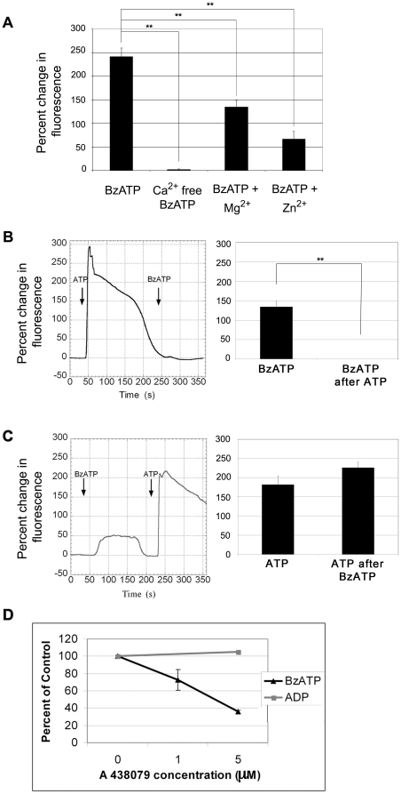
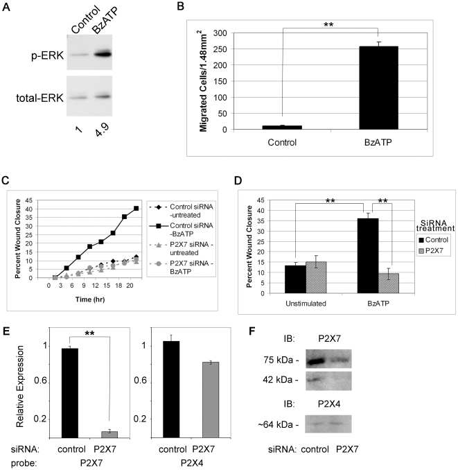
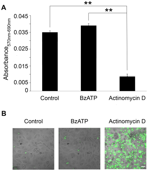
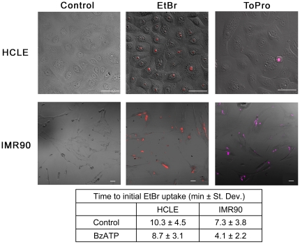
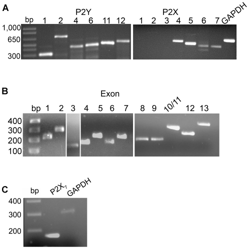
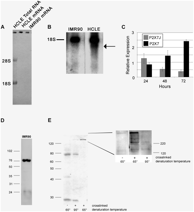
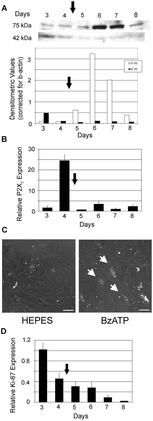
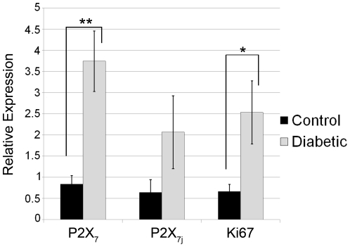
Improper wound repair of the corneal epithelium can alter refraction of light resulting in impaired vision. We have shown that ATP is released after injury, activates purinergic receptor signaling pathways and plays a major role in wound closure. In many cells or tissues, ATP activates P2X(7) receptors leading to cation fluxes and cytotoxicity. The corneal epithelium is an excellent model to study the expression of both the full-length P2X(7) form (defined as the canonical receptor) and its truncated forms. When Ca(2+) mobilization is induced by BzATP, a P2X(7) agonist, it is attenuated in the presence of extracellular Mg(2+) or Zn(2+), negligible in the absence of extracellular Ca(2+), and inhibited by the competitive P2X7 receptor inhibitor, A438079. BzATP enhanced phosphorylation of ERK. Together these responses indicate the presence of a canonical or full-length P2X(7) receptor. In addition BzATP enhanced epithelial cell migration, and transfection with siRNA to the P2X(7) receptor reduced cell migration. Furthermore, sustained activation did not induce dye uptake indicating the presence of truncated or variant forms that lack the ability to form large pores. Reverse transcription-polymerase chain reaction and Northern blot analysis revealed a P2X(7) splice variant. Western blots identified a full-length and truncated form, and the expression pattern changed as cultures progressed from monolayer to stratified. Cross-linking gels demonstrated the presence of homo- and heterotrimers. We examined epithelium from age matched diabetic and non-diabetic corneas patients and detected a 4-fold increase in P2X(7) mRNA from diabetic corneal epithelium compared to non-diabetic controls and an increased trend in expression of P2X(7)variant mRNA. Taken together, these data indicate that corneal epithelial cells express full-length and truncated forms of P2X(7), which ultimately allows P2X(7) to function as a multifaceted receptor that can mediate cell proliferation and migration or cell death.


Conflict of interest statement

Competing Interests: The authors have declared that no competing interests exist.

Figures

Figure 1
Figure 1. BzATP causes an increase in intracellular Ca2+.
HCLE cells were incubated in 5 µM fluo-3AM for 30 min and imaged in a flow-through apparatus on a Zeiss LSM 510 confocal microscope. Cells were washed with indicated HEPES-buffered saline (control, Ca2+ free, 4 mM Mg2+, 100 µM ZnSO4) and stimulated with BzATP in corresponding buffer for 2 min. Maximal percent change in average fluorescence of a 460 µm×460 µm field was determined. A. HCLE cells stimulated with BzATP in HEPES buffer, in Ca2+ free - HEPES buffer, in HEPES buffer with Mg2+ and in HEPES buffer with Zn2+. Graphs represent a minimum of six independent experiments +/− SEM. Analysis of variance was determined using the general linear model procedure followed by Tukey-Kramer posthoc test. **p<0.001. B. Response of HCLE cells to ATP followed by stimulation with BzATP. Representative trace of changes in fluorescence over time. Graph represents difference between stimulation with ATP and BzATP after ATP and represents a minimum of six independent experiments +/− SEM. Students t-test ** p<0.001. C Response of HCLE cells to BzATP followed by ATP. Representative trace of changes in fluoresence over time. Graph represents difference between stimulation with BzATP and ATP after BzATP and represents a minimum of six independent experiments +/− SEM. D. Cells were washed with HEPES buffered saline with the indicated concentrations of A438079 and stimulated with BzATP or equimolar concentrations of ADP for 2 min in the presence or absence of inhibitor. The average maximum percent change in fluorescence is graphed as a percentage of the uninhibited control +/− SEM. Each time point is an average of 12 independent experiments. Linear regression analysis where the slope of the line for BzATP is significantly different from 0. p = 0.0001. The control ADP is not significantly different from 0.
Figure 2
Figure 2. P2X7 activation induces phosphorylation of ERK and cell migration.
A. Cells were treated in the presence or absence of BzATP for 5 min, lysed and equivalent amounts of protein were resolved by SDS-PAGE and immunoblotted with anti-p-ERK. Blots were stripped and reprobed with anti-ERK and pERK normalized to ERK. Significance was determined by Student's test (p<0.05). Blot is representative of 3 independent experiments. B. Transwell migration assays were performed for 8 hr in the presence of BzATP or binding buffer (control). Migrated cells were stained with propidium iodide, counted in 6 randomly chosen fields (1.48 mm2) and averaged. Data are representative of 3 independent experiments and are presented as mean +/− SEM. Significance was determined by Student's t-test: **p<0.001. C. Downregulation of P2X7 attenuates wound healing in a representative experiment. HCLE cells transfected with control (non-targeting siRNA) or siRNA targeted to P2X7 receptor were cultured to confluence. Unsupplemented media or media with 100 µM BzATP were added prior to injury. Slides were placed on a heated stage in an environmental chamber at 37°C and 5% CO2. Scratch wounds were made and contiguous images were taken every 20 min over 20 hr. Data are representative of 3 independent experiments. D. Percent wound closure from directed migration experiments at endpoint are presented as mean +/− SEM. Significance was determined by ANOVA followed by Tukey posthoc test: **p<0.001. E. HCLE cells transfected with control (non-targeting siRNA) or siRNA targeted to P2X7 receptor were cultured to confluence. Real time RT-PCR was performed and relative expression of the indicated receptors was determined using the ΔΔCT method. The average relative expression of 3 independent experiments +/− SEM is presented. Significance was determined by Student's test: **p<0.004. F. Parallel experiments to E. were conducted and cultures were lysed and equivalent amounts of protein resolved by SDS-PAGE and immunoblotted with antibodies directed to P2X7 and P2X4 receptors.
Figure 3
Figure 3. Cytotoxicity is not induced in epithelial cells following P2X7 activation.
A. MTT assay for cell toxicity was performed on confluent HCLE cells. The response to control media, 100 µM BzATP or actinomycin D is presented as corrected absorbance (570nm–690nm). Data are representative of 3 independent experiments and are presented as +/− SEM. (Significance was determined by ANOVA followed by Tukey posthoc test; ** p<0.001. B. HCLE cells were stimulated with 100 µM BzATP, 2 µg/ml actinomycin D or control media lacking growth factors for 20 hr. Caspase activation was detected with NucView 488 Live Cell Caspase-3 Assay (scale bar  = 50 µm and applies to all 3 images). Images are representative of 5 independent experiments.
Figure 4
Figure 4. Confluent monolayer corneal epithelial cells do not form large pores when stimulated with BzATP.
HCLE or IMR90 cells were cultured and stimulated with control media or media containing 100 µM BzATP in the presence of EtBr or ToPro-3 and imaged over 20 min while maintained at 37°C and 5% CO2 in an environmental chamber on a Zeiss LSM 510 confocal microscope. Representative images are of cells after 20 min BzATP with EtBr (EtBr) or BzATP with ToPro-3 (ToPro)(Scale bar  = 50 µm). Control treated cells in the presence of EtBr are shown (Control). ToPro-3 is detected in IMR90 cells and only detected in HCLE cells being shed. The time to initial EtBr uptake is presented. Images are representative of 3 independent experiments.
Figure 5
Figure 5. Expression of receptor transcripts in epithelial cells.
A. RT-PCR was performed on HCLE mRNA showing presence of P2Y 1, 2, 4, 6, 11, and 12 receptor transcripts and P2X 4, 5, 6, and 7 receptor transcripts. GAPDH is included as internal control. B. PCR was performed on genomic DNA using exon specific primers. C. RT-PCR was performed on HCLE mRNA using primers that spanned exon 8 (to amplify variants f and j), showing an expected product size of 169 bp. GAPDH was amplified as an internal control. All products were sequenced and verified. Data is representative of a minimum of 3 independent experiments.
Figure 6
Figure 6. HCLEs express a full length and truncated P2X7 mRNA transcript and protein.
A. mRNA was purified from total RNA of HCLE and IMR90 cells cultured for 72 hours by twice passing over an oligo-dT column. Purity was confirmed by methylene blue staining of nytran filter following transfer (note lack of rRNA in mRNA lanes). B. Northern blot analysis of HCLE and IMR90 mRNA from 72 hour cultures. The probe used was generated by PCR amplification of a consensus region in all P2X7 variants. The relative position of 18s rRNA is indicated and the smaller transcript expressed by epithelial cells is indicated (arrow). C. Expression of P2X7 and P2X7j mRNA transcript over a 72 hour incubation. Relative expression was determined using the ΔΔCT method. D. Expression of P2X7 receptor by IMR90 cells. Cells were cultured for 72 hours, lysed and protein was resolved by SDS-PAGE (12%) and immunoblotted with anti-P2X7. E. Expression of P2X7 receptor by HCLE cells. Cells were cultured for 72 hours, lysed and protein was resolved by SDS-PAGE (12%) and immunoblotted with anti-P2X7. Hetero- and homotrimers are identified on crosslinking gels. Epithelial cells were cultured for 72 hours, incubated in situ in the presence of formaldehyde, lysed and incubated at 65°C or 95°C to maintain or break crosslinks. Inset – equivalent experiment resolved by SDS-PAGE (8%) and immunoblotted with anti-P2X7. Data is representative of a minimum of 3 experiments.
Figure 7
Figure 7. Expression of P2X7 receptor changes with stratification.
A. Stratification was induced after day 4 (arrow) and cells were cultured for 4 additional days. Cells were lysed and equivalent amounts of protein were resolved by SDS-PAGE and immunoblotted with anti-P2X7. Representative blot is shown. Quantification of 75 kDa and 42 kDa protein are graphed as densitometric values relative to β-actin. Data is representative of a minimum of 4 independent experiments. B. Relative expression of P2X7 mRNA was determined using the ΔΔCT method and the average of 3 independent cultures +/− SEM is presented. C. Apical cells in stratified culture of HCLE cells at day 7 prestimulated with BzATP show minimal uptake of ToPro-3 using the flow through aparatus. Cultures were prestimulated with BzATP for 5 minutes at which time ToPro-3 in the presence of BzATP was added. Cells were imaged for an additional 20 minutes in the presence of BzATP as described in Figure 4 (scale bar  = 50 µm). Arrow indicates representative areas of uptake. Control experiments were performed in the presence of HEPES buffer. D. Relative expression of Ki67 mRNA was determined using the ΔΔCT method and the average of 3 independent cultures +/− SEM is presented.
Figure 8
Figure 8. Diabetic corneal epithelium displays enhanced P2X7 receptor expression compared to control non-diabetic corneal epithelium.
Central corneal epithelium from 6 samples of control and diabetic were analyzed using real time PCR. Relative expression of the P2X7 receptor (all variants), P2X7j variant receptor, and Ki67 were determined using the ΔΔCT method. The average relative expression of 6 independent samples +/− SEM is presented. Significance was determined by Student's t-test: **p<0.002, *p<0.05 respectively.

Similar articles

Cited by

References

    1. Abbracchio MP, Burnstock G. Purinergic Signaling: Pathophysiologic Roles. Jpn J Pharmacol. 1998;78:113–145. - PubMed
    1. Ralevic V, Burnstock G. Receptors for Purines and Pyrimidines. Pharmacol Rev. 1998;50:413–492. - PubMed
    1. Weinger I, Klepeis V, Trinkaus-Randall V. Tri-nucleotide receptors play a critical role in epithelial cell wound repair. Purinergic Signalling B. 2005. pp. 281–292. - PMC - PubMed
    1. North RA. Molecular Physiology of P2X Receptors. Physiol Rev. 2002;82:1013–1067. - PubMed
    1. Ferrari D, Pizzirani C, Adinolfi E, Lemoli RM, Curti A, et al. The P2X7 Receptor: A key player in IL-1 processing and release. J Immunol. 2006;176:3877–3883. - PubMed

Publication types

MeSH terms