. 2011;6(12):e28546.
doi: 10.1371/journal.pone.0028546. Epub 2011 Dec 5.

Characterization of a new mouse model for peripheral T cell lymphoma in humans

Affiliations

Characterization of a new mouse model for peripheral T cell lymphoma in humans

Niklas Beyersdorf et al. PLoS One. 2011.

Abstract

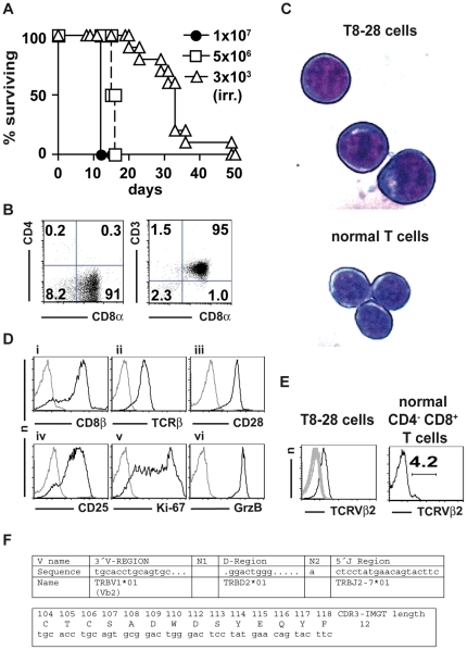
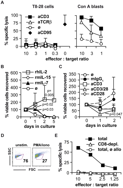
Peripheral T cell lymphomas (PTCLs) are associated with a poor prognosis due to often advanced disease at the time of diagnosis and due to a lack of efficient therapeutic options. Therefore, appropriate animal models of PTCL are vital to improve clinical management of this disease. Here, we describe a monoclonal CD8(+) CD4(-) αβ T cell receptor Vβ2(+) CD28(+) T cell lymphoma line, termed T8-28. T8-28 cells were isolated from an un-manipulated adult BALB/c mouse housed under standard pathogen-free conditions. T8-28 cells induced terminal malignancy upon adoptive transfer into syngeneic BALB/c mice. Despite intracellular expression of the cytotoxic T cell differentiation marker granzyme B, T8-28 cells appeared to be defective with respect to cytotoxic activity as read-out in vitro. Among the protocols tested, only addition of interleukin 2 in vitro could partially compensate for the in vivo micro-milieu in promoting growth of the T8-28 lymphoma cells.


Conflict of interest statement

Competing Interests: The authors have declared that no competing interests exist.

Figures

Figure 1
Figure 1. In vivo tumorigenicity of and T cell marker expression by T8-28 cells.
(A) Survival of unmanipulated BALB/c.OlaHsd mice transplanted with 1×10⁷ (experiment 1; n = 2) or 5×10⁶ (experiment 2; n = 2) T8-28 cells or irradiated BALB/c.OlaHsd mice transplanted with 3×10³ T8-28 cells on day 0 (pooled data of two experiments; n = 10). (B) CD4, CD8α and CD3 expression by T8-28 cells. Figures indicate the percentages of cells in the respective quadrants. (C) Cytospins of T8-28 and freshly isolated mouse T cells stained by the Diff-Quik® method. Original magnification was 630-fold. Microscope: LEICA DMIRE2. Camera: LEICA DFC300 FX. Acquisition Software: LEICA IM50 Image Manager. Adobe Photoshop CS3 was used to obtain the close-ups shown. (D) Fluorescence-activated cell sorting analysis of T cell lineage and activation markers as indicated (black histograms). Staining controls (grey histograms): i, v, isotype-matched control mAb; ii,vi, non-isotype-matched control mAb; iii, no primary mAb; iv: identical control staining as in iii. (E) Expression of TCRVβ chains by T8-28 cells and CD4 CD8+ T cells of a normal BALB/c mouse as indicated. Grey histograms represent overlays of the 14 remaining TCRVβ chains (Vβ3–Vβ18) detected by the kit used. (F) The genetic setup and primary structure of the TCR CDR3 region was analyzed by polymerase chain reaction DNA sequencing and the IMGT/V-QUEST program. Please note that TRBV1 in the IMGT nomenclature is identical to designation as Vβ2 used throughout this paper and that the *01 allele has originally been found in BALB/c mice which is consistent with the origin of the T8-28 cells. The sequenced Cβ part (not shown) could be identified as Cβ2 as expected for a TCR-chain using the second DJ cluster.
Figure 2
Figure 2. T cell effector functions of T8-28 cells and susceptibility to killing by allogeneic T cells.
(A) T8-28 cells (left) or Con A blasts (right) were used as effector cells in re-directed lysis assays against A20J cells in the presence or absence of the indicated antibodies. Means ± SD of three to six independent experiments are shown. (B) Freshly thawed T8-28 cells were cultured either alone or in the presence of 10−6 M of the indicated cytokines. Means ± SD of two to five experiments are shown. (C) Cell recovery after culture of T8-28 cells in the presence of 10−6 M rhIL-2 and paramagnetic beads coated with anti-CD3 and/or anti-CD28 mAbs as indicated. P-values in (B) and (C) are the results of two-way ANOVA testing against the cells without the specified treatment (‘ø’). (D) Percentage of viable cells as determined by FSC/SSC characteristics among un-stimulated and PMA/ionomycin re-stimulated T8-28 cells. The figures indicate the number of cells within the life gate. The data are representative for more than three experiments with comparable results. (E) T8-28 cells are susceptible to killing by allogeneic CD8+ T cells. CD8+ cell-depleted allogeneic lymph node cells (triangles) and lymph node cells cultured without allogeneic APCs (circles) were used as controls. Means of duplicates are shown. The experiment was repeated with similar result.

References

    1. Mulloy JC. Peripheral T cell lymphoma: new model + new insight. J Exp Med. 2010;207:911–913. - PMC - PubMed
    1. van Hall T, van Bergen J, van Veelen PA, Kraakman M, Heukamp LC, et al. Identification of a novel tumor-specific CTL epitope presented by RMA, EL-4, and MBL-2 lymphomas reveals their common origin. J Immunol. 2000;165:869–877. - PubMed
    1. Gorer PA. Studies in antibody response of mice to tumour inoculation. Br J Cancer. 1950;4:372–379. - PMC - PubMed
    1. Pechloff K, Holch J, Ferch U, Schweneker M, Brunner K, et al. The fusion kinase ITK-SYK mimics a T cell receptor signal and drives oncogenesis in conditional mouse models of peripheral T cell lymphoma. J Exp Med. 2010;207:1031–1044. - PMC - PubMed
    1. Dierks C, Adrian F, Fisch P, Ma H, Maurer H, et al. The ITK-SYK fusion oncogene induces a T-cell lymphoproliferative disease in mice mimicking human disease. Cancer Res. 2010;70:6193–6204. - PubMed

Publication types

MeSH terms