. 2011;6(12):e28587.
doi: 10.1371/journal.pone.0028587. Epub 2011 Dec 6.

Tyrosine phosphorylation of Rac1: a role in regulation of cell spreading

Affiliations

Tyrosine phosphorylation of Rac1: a role in regulation of cell spreading

Fumin Chang et al. PLoS One. 2011.

Abstract

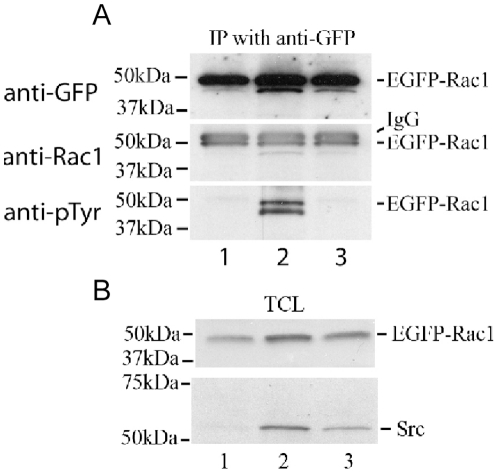
Rac1 influences a multiplicity of vital cellular- and tissue-level control functions, making it an important candidate for targeted therapeutics. The activity of the Rho family member Cdc42 has been shown to be modulated by tyrosine phosphorylation at position 64. We therefore investigated consequences of the point mutations Y64F and Y64D in Rac1. Both mutations altered cell spreading from baseline in the settings of wild type, constitutively active, or dominant negative Rac1 expression, and were accompanied by differences in Rac1 targeting to focal adhesions. Rac1-Y64F displayed increased GTP-binding, increased association with βPIX, and reduced binding with RhoGDI as compared with wild type Rac1. Rac1-Y64D had less binding to PAK than Rac1-WT or Rac1-64F. In vitro assays demonstrated that Y64 in Rac1 is a target for FAK and Src. Taken together, these data suggest a mechanism for the regulation of Rac1 activity by non-receptor tyrosine kinases, with consequences for membrane extension.


Conflict of interest statement

Competing Interests: The authors have declared that no competing interests exist.

Figures

Figure 1
Figure 1. Sequence homology for Cdc42 and Rac1.
Amino acid sequences are shown for human Cdc42 (top) and Rac1 (bottom). The sequence identity between the two proteins at aa 61–70 is underlined, Y64 is capitalized, and all tyrosine residues appear in bold type.
Figure 2
Figure 2. Rac1-Y64 phosphorylation modulated cell spreading.
(A) Transfected HUVEC were plated onto fibronectin-coated coverslips for 90 minutes and then fixed and permeabilized for immunofluorescence staining. Rhodamine-phalloidin labeled EGFP-positive cells were used for cell surface area quantification using Openlab software. Data are shown as mean±SEM, and were as follows: non-transfected HUVEC controls had a mean surface area of 7760 µm2 (± 730.5, n = 28), and the mean area was 6510 µm2 (±1433.0, n = 8) for EGFP-transfected cells. A large increase in spread surface area to 10246 µm2 (±1310, n = 14) was noted in EGFP-Rac1-Y64F-transfected HUVEC, and cells expressing EGFP-Rac1-Y64D had a greatly reduced mean surface area of 1480 µm2 (±1037, n = 5). ** indicates p <0.001, and *** denotes p < 0.0001 for the designated population as compared with the EGFP control. (B) Cell surface area was quantified from phalloidin-labeled MEF images using Openlab software in cells transfected with EGFP-Rac1-WT, EGFP-Rac1-Y64F, EGFP-Rac1-Y64D, EGFP-Rac1-61L, EGFP-Rac1-61L/64F, EGFP-Rac1-61L/64D, EGFP-Rac1-17N, EGFP-Rac1-17N/64F, or EGFP-Rac1-17N/64D. Data are shown as the mean±SEM. * denotes p < 0.05, ** means p <0.001, and *** is p < 0.0001 when data from groups transfected with Y64F or Y64D are compared with values from the corresponding controls (e.g. EGFP-Rac1-WT, EGFP-61L, or EGFP-17N, respectively, without the mutations at position 64). ## denotes p<0.005, while ### denotes p<0.0001 when data from 61L or 17N mutants were compared with wild-type. Quantitative cell spreading data and the number of MEF studied in each group are detailed in Table 1. (C) MEF were transfected with EGFP, or EGFP-tagged Rac1 single mutants. Two days after transfection, cells were seeded onto fibronectin-coated coverslips for two hours and prepared for immunofluorescence analysis. Cells were labeled with DAPI, rhodamine-phalloidin and anti-vinculin (followed by Cy5). Transfected GFP-positive cells are shown indicated in black and white, while the composite images display merges of combined DAPI (blue), phalloidin (red) and vinculin (green) labeling. Scale bar denotes 20 µm. (D) MEF that had been transfected with the EGFP-tagged Rac1 double mutants including Rac1-61L/64F, -61L/64D, -17N/64F, or -17N/64D were plated on fibronectin-coated coverslips and labeled for IF analysis as describe for panel 2C. Scale is the same as panel 2C.
Figure 3
Figure 3. Expression of Rac1-Y64F or Rac1-Y64D altered lamellipodial dynamics.
HUVEC cells were transfected with EGFP, or EGFP-tagged Rac1-Y64D, or Rac1-Y64F. Cells were plated onto fibronectin-coated coverslips two days after transfection, and were observed under phase contrast microscopy until initial attachment was achieved (typically 10 minutes). Images were taken every 20 seconds for 20 minutes. Kymograph analysis of cell spreading efficiency was performed using these serial images. (A) Three sets of kymographs are shown from each transfected cell: a–c for EGFP-Rac1-Y64F; d-f for EGFP and g-i for EGFP-Rac1-Y64D. The lower panel shows the transfected cells that were studied with the line scan used for kymograph. The inset shows the GFP signal of the transfected cells. (B) Kymography from three to six transfected cells were analyzed to study the differences in their spreading. Significant differences in lamellipodial extension were noted in both cells expressing Rac1-Y64F and Rac1-Y64D when compared with HUVEC expressing Rac1-WT (p values  =  0.0377 and 0.0005 respectively).
Figure 4
Figure 4. Y64 phosphorylation modulated focal adhesion targeting of active Rac1.
(A) HUVEC cells were transfected with EGFP-tagged Rac1 mutants including Rac1-61L, Rac1-61L64F and Rac1-61L64D. Cells were labeled with DAPI and anti-vinculin (followed by Cy5). Transfected EGFP-positive cells are shown in green, while the vinculin labeled focal adhesions shown in red. The composite images display merges of combined GFP (green), DAPI (white), and vinculin (red) labeling. The boxed areas were magnified (2X) and shown in the insets. Scale bar denotes 30 µm. (B) The percentage of the total focal adhesion area (defined by vinculin labeling) that was occupied by EGFP-tagged Rac1 mutants in transfected cells were quantified using Openlab software. Fifteen randomly picked transfected cells were analyzed in each group, and data are shown as the mean±SD. * denotes p < 0.05 when data from groups transfected with 61L/64F or 61L/64D are compared with values from the group transfected with 61L alone.
Figure 5
Figure 5. Mutations in Rac1-Y64 changed binding to GTP.
(A) Left panel: MEF cells were transfected with Rac1 mutants including EGFP-Rac1-61L (lane 1 EGFP-Rac1-17N (lane 2), EGFP-Rac1-Y64D (lane 3), or EGFP-Rac1-Y64F (lane 4), or with EGFP (lane 5), or EGFP-Rac1-WT (lane 6). Coomassie blue staining of the membrane (bottom row, left) verified the addition of an equal amount of PBD-GST. Western blotting was done using both anti-Rac1 and anti-GFP antibodies. Right panel: 15 µg of total protein lysates were also analyzed by SDS-PAGE and immunoblotted to verify equal loading and the expression of the transfected EGFP-Rac1 constructs. (B) Four sets of Rac1 activity assay results were used for densitometry and statistical analysis of the difference in activity between Rac1-WT and Rac1-Y64F. Western blots were scanned and Rac1 activity was normalized to the total cellular Rac1. Data shown are mean values±SEM. * denotes p < 0.02. (C) Protein lysates were harvested from MEF (lane 1) or MEF that were transfected with EGFP-Rac1WT (lane 2), EGFP-Rac1-61L (lane 3), EGFP-Rac1-61L/64F (lane 4), or EGFP-Rac1-61L/64D (lane 5). PBD-GST pull down assays were performed with 800 µg of total protein to determine Rac1 activity. The membrane was immunoblotted with anti-Rac1 to demonstrate the level of Rac1 activity, and was then stained with Coomassie blue to ascertain the relative amounts of PBD-GST substrate added to each sample. Expression efficiency of the wild type and mutant EGFP-Rac1 constructs was assayed using 15 µg of total protein from whole cell lysates and the Rac1 activity was normalized to the respective protein level of each sample. (D) Five sets of experimental data were analyzed to examine the differences between the Rac1 activity in lysates from MEF expressing EGFP-RacWT, EGFP-Rac1-61L, EGFP-Rac1-61L/64F, and EGFP-Rac1-61L/64D. ECL blots were scanned and analyzed using Image J software. The intensity of each of the active Rac1 bands pulled down by PBD was normalized to the total cellular Rac protein band and calculated by a software-based algorithm. The Rac1 activity in lysates from cells expressing EGFP-Rac1-61L was assigned a relative value of 100%. ## indicated a significant decrease of Rac1 activity in 61L/64D compared with 61L (p < 0.01). * indicated a significant increase in Rac1 activity in 61L, 61L/64F and 61L/64D compared with Rac1 wild-type (p > 0.001).
Figure 6
Figure 6. The Y64F mutation in Rac1 changed binding to guanine nucleotide exchange factors.
(A) Protein lysates were harvested from MEF that were co-transfected with Flag-tagged βPIX, and either EGFP-tagged Rac1WT (lane 1) or Rac1-Y64F (lane 2). Ectopically expressed EGFP-Rac1 proteins, wild-type or Y64F mutant, were immunoprecipitated with anti-GFP antibody and the membrane was blotted with anti GFP to demonstrate the expression level of EGFP-Rac1 proteins and also with anti-Flag antibody to reveal the Flag-βPIX that was co-immunoprecipitated with EGFP-Rac1 proteins. Immunoblotting of total cell lysates (TCL) for Flag-βPIX and EGFP-Rac1 are shown below the co-immunoprecipitation study as controls. (B) Four sets of experimental data were analyzed for the differences in binding to Flag-tagged βPIX for EGFP-Rac1WT or EGFP-Rac1-Y64F. The intensity of the co-immunoprecipitated Flag-βPIX was normalized to the EGFP-Rac1 pulled down by anti-GFP antibody. The amount of Flag-βPIX co-immunoprecipitated with EGFP-Rac1WT was assigned a relative value of 100%. EGFP-Rac1-Y64F bound more efficiently to Flag-βPIX (p  =  0.0223). (C) Protein lysates were harvested from MEF that were co-transfected with Myc-tagged Tiam1 and either EGFP-Rac1WT (lane 1) or EGFP-Rac1-Y64F (lane 2). Ectopically expressed EGFP-Rac1 proteins were immunoprecipitated with anti-GFP antibody and the membrane was blotted with anti GFP to demonstrate the expression level of EGFP-Rac1 proteins, and with anti-Myc antibody to reveal the Myc-Tiam1 that was co-immunoprecipitated with the EGFP-Rac1 constructs. Immunoblotting of total cell lysates (TCL) for Myc-Tiam1 and EGFP-Rac1 are shown below the co-immunoprecipitation study as controls. (D) Six sets of experimental data were analyzed for differences in binding to Myc-Tiam1 between EGFP-Rac1WT and EGFP-Rac1-Y64F. The intensity of the immunoprecipitated Myc-Tiam1 was normalized to the EGFP-Rac1 pulled down by anti-GFP antibody. The amount of Myc-Tiam1 co-immunoprecipitated with EGFP-Rac1WT was assigned a relative value of 100%. The difference did not reach statistical significance p  =  0.3793).
Figure 7
Figure 7. The Rac1-64D mutation caused decreased Rac1 binding to PAK.
30 µg of purified GST-tagged Rac1 17N (lane 1), wild-type (lane 2), 61L (lane 3), 64D (lane 4) or 64F (lane 5) were bound to sepharose beads and loaded with GTPγS for 30 min before incubation with 400 µg of HUVEC protein lysates in order to assay PAK binding. Unlike the 17N mutant which can not bind GTP, Rac1-64D does bind GTP and pull down PAK. However, PAK binding by this mutant is reduced compared to that seen for Rac1-WT, -61L, or -64F.
Figure 8
Figure 8. The Y64F mutation in Rac1 decreased binding to RhoGDI.
(A) Protein lysates were harvested from MEF that were co-transfected with Myc-tagged RhoGDI and either EGFP-tagged Rac1WT (lane 1) or Rac1-Y64F (lane 2). EGFP-Rac1WT or EGFP-Rac1-Y64F proteins were immunoprecipitated with anti-GFP antibody and the membrane was blotted first with anti-GFP to demonstrate the expression level of EGFP-Rac1 proteins, and then with anti-Myc antibody to show the Myc-RhoGDI that co-immunoprecipitated with EGFP-Rac1 proteins. Immunoblotting of total cell lysates (TCL) for Myc-RhoGDI and EGFP-Rac1 are shown below the co-immunoprecipitation study as controls. (B) Five sets of experimental data were analyzed for the differences between EGFP-Rac1WT and EGFP-Rac1-Y64F interaction with Myc-RhoGDI. ECL blots were scanned, normalized, and analyzed as described above. Myc-RhoGDI bound less well to EGFP-Rac1-Y64F as compared to EGFP-Rac1WT (p  =  0.011).
Figure 9
Figure 9. Src and FAK mediated tyrosine phosphorylation of Rac1 at tyrosine 64 in vitro.
(A) 5 µg of either GST-Rac1 (lanes 1–5) or GST (lane 6) were incubated with 0 µg (lane1), 0.25 µg (lane 2), 1 µg (lanes 3, 5, 6), or 4 µg (lane 4) of purified Src in kinase buffer. All samples included 20 µM ATP except for the one shown in lane 5. The kinase reaction mixture was analyzed by SDS-PAGE and immunoblotted with antibodies against phosphotyrosine, Src, and GST. (B) 5 µg of GST-Rac1 (lanes 1–4) or GST (lanes 5–8) were incubated with 0 µg (lanes 4, 8), 0.5 µg (lanes 1, 5), 1.5 µg (lanes 2, 6), or 2.5 µg (lanes 3, 7) of GST-tagged wild-type FAK (GST-wtFAK411-686) in kinase buffer with 20 µM ATP. The kinase reaction mixture was analyzed by SDS-PAGE, and immunoblotted with antibodies against phosphotyrosine and GST. (C) 5 µg of GST-Rac1-WT (lanes 1–4) or GST-Rac1-Y64F (lanes 5–8) were incubated with 0 µg (lanes 1, 5), 1 µg (lanes 2, 6), or 3 µg (lanes 3, 4, 7, 8) of purified Src. Reactions run in all lanes contained 20 µM ATP, except for lanes 4 and 8. The kinase reaction mixture was analyzed by SDS-PAGE and immunoblotted with antibodies against phosphotyrosine, Src, and GST. (D) 5 µg of GST-Rac1-WT (lanes 1–4) or GST-Rac1-Y64F (lanes 5–8) were incubated with purified GST-WT-FAK (aa 411-686) in the following amounts: 0 µg (lanes 1, 5), 1 µg (lanes 2, 6), or 3 µg (lanes 3, 4, 7, 8). All reactions included 20 µM ATP except for the ones represented in lanes 4 and 8. The kinase reaction mixture was analyzed by SDS-PAGE and immunoblotted with antibodies against phosphotyrosine and GST.
Figure 10
Figure 10. Src tyrosine-phosphorylated Rac1 in MEF.
(A) SYF cells were transfected with EGFP- Rac1-WT alone (lane 1) or together with wild-type Src (lane 2) or kinase dead Src (lane 3). EGFP-Rac1 immunoprecipitates were obtained with anti-GFP antibody two days after transfection, and immunoblotted with anti-GFP and anti-Rac1antibodies to quantify EGFP-Rac1 expression, and then with anti-phosphotyrosine to probe for tyrosine phosphorylation of EGFP-Rac1, as seen in cells that were transfected with exogenous Src. Molecular weight markers are shown in kilodaltons (kDa) on the left. (B) Immunoblotting of total cell lysates (TCL) for EGFP-Rac1 and Src are shown as a control.

Similar articles

Cited by

References

    1. Bosco EE, Mulloy JC, Zheng Y. Rac1 GTPase: a "Rac" of all trades. Cell Mol Life Sci. 2009;66:370–374. - PMC - PubMed
    1. Briancon-Marjollet A, Ghogha A, Nawabi H, Triki I, Auziol C, et al. Trio mediates netrin-1-induced Rac1 activation in axon outgrowth and guidance. Mol Cell Biol. 2008;28:2314–2323. - PMC - PubMed
    1. Hordijk PL. Regulation of NADPH oxidases: the role of Rac proteins. Circ Res. 2006;98:453–462. - PubMed
    1. Kissil JL, Walmsley MJ, Hanlon L, Haigis KM, Bender Kim CF, et al. Requirement for Rac1 in a K-ras induced lung cancer in the mouse. Cancer Res. 2007;67:8089–8094. - PubMed
    1. Nikolova E, Mitev V, Zhelev N, Deroanne CF, Poumay Y. The small Rho GTPase Rac1 controls normal human dermal fibroblasts proliferation with phosphorylation of the oncoprotein c-myc. Biochem Biophys Res Commun. 2007;359:834–839. - PubMed

Publication types

MeSH terms