. 2011;6(12):e28591.
doi: 10.1371/journal.pone.0028591. Epub 2011 Dec 6.

High ACSL5 transcript levels associate with systemic lupus erythematosus and apoptosis in Jurkat T lymphocytes and peripheral blood cells

Affiliations

High ACSL5 transcript levels associate with systemic lupus erythematosus and apoptosis in Jurkat T lymphocytes and peripheral blood cells

Antonio Catalá-Rabasa et al. PLoS One. 2011.

Abstract

Background: Systemic lupus erythematosus (SLE) is a prototypical autoimmune disease in which increased apoptosis and decreased apoptotic cells removal has been described as most relevant in the pathogenesis. Long-chain acyl-coenzyme A synthetases (ACSLs) have been involved in the immunological dysfunction of mouse models of lupus-like autoimmunity and apoptosis in different in vitro cell systems. The aim of this work was to assess among the ACSL isoforms the involvement of ACSL2, ACSL4 and ACSL5 in SLE pathogenesis.

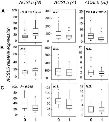
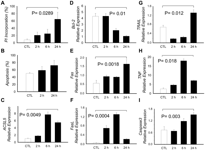
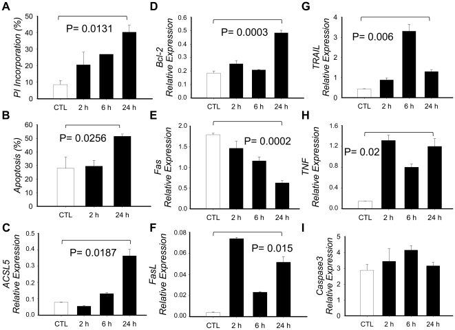
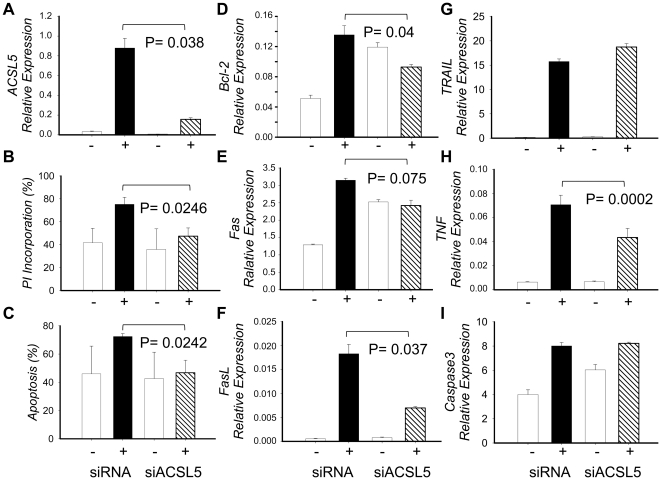
Findings: With this end, we determined the ACSL2, ACSL4 and ACSL5 transcript levels in peripheral blood mononuclear cells (PBMCs) of 45 SLE patients and 49 healthy controls by quantitative real time-PCR (q-PCR). We found that patients with SLE had higher ACSL5 transcript levels than healthy controls [median (range), healthy controls = 16.5 (12.3-18.0) vs. SLE = 26.5 (17.8-41.7), P = 3.9×10 E-5] but no differences were found for ACSL2 and ACSL4. In in vitro experiments, ACSL5 mRNA expression was greatly increased when inducing apoptosis in Jurkat T cells and PBMCs by Phorbol-Myristate-Acetate plus Ionomycin (PMA+Io). On the other hand, short interference RNA (siRNA)-mediated silencing of ACSL5 decreased induced apoptosis in Jurkat T cells up to the control levels as well as decreased mRNA expression of FAS, FASLG and TNF.

Conclusions: These findings indicate that ACSL5 may play a role in the apoptosis that takes place in SLE. Our results point to ACSL5 as a potential novel functional marker of pathogenesis and a possible therapeutic target in SLE.


Conflict of interest statement

Competing Interests: The authors have declared that no competing interests exist.

Figures

Figure 1
Figure 1. Association of ACSL5 mRNA expression levels with SLE.
A) ACSL5 transcript levels in PBMCs from healthy controls (0) and SLE patients (1) for ACSL5(N) (number of samples n: 0 = 49, 1 = 45), activated ACSL5(A) (n, 0 = 34, 1 = 40) and the ratio A/N (ACSL5(SI) (n, 0 = 34, 1 = 40). B) ACSL5 transcript levels in PBMCs from males (0) (n = 24) and females (1) (n = 25), all of them from the control group. C) ACSL5 transcript levels in PBMCs from prednisone treated (1) (n = 28) and untreated (0) (n = 12) SLE patients. Results are represented in box plots given medians, quartiles, outsiders (circle points) and extremes (asterisk points). P-values of differences between the groups (0) and (1) are defined by Mann-Whitney Statistics. N.S. stands for non significant.
Figure 2
Figure 2. Effect of PMA+Io activation in PBMCs.
PBMCs from healthy controls were obtained and cultured in the presence or absence of PMA+Io for up to 24 h. A) Cells were collected at defined times, washed with PBS, fixed with 70% Ethanol, stained with Propidium Iodide (PI) and analyzed in a FACSCaliburTM flow cytometer to determine the apoptotic hipodiploid cell fragments (defined as percentage of PI incorporation). B) Cells were washed twice with PBS and double stained with Annexin V and PI, then analyzed by FACS to determine the percentage of apoptotic cells (Annexin V positive and double positive cells). C–I) Total RNA was extracted, cDNA synthesized and qRT-PCR implemented to determine mRNA expression. Results are given by means of three independent experiments and the bars show the standard deviation. P-value has been calculated with the paired Student t test.
Figure 3
Figure 3. Effect of PMA+Io activation in Jurkat T cells.
Jurkat T cells were cultured in the presence or absence of PMA+Io for up to 24 h. A) Cells were collected at defined times, washed with PBS, fixed with 70% Ethanol, stained with Propidium Iodide (PI) and analyzed in a FACSCaliburTM flow cytometer to determine the apoptotic hipodiploid cell fragments (defined as percentage of PI incorporation). B) Cells were washed twice with PBS and double stained with Annexin V and PI, then analyzed by FACS to determine the percentage of apoptotic cells (Annexin V positive and double positive cells). C–I) Total RNA was extracted, cDNA synthesized and qRT-PCR implemented to determine mRNA expression. Results are given by means of three independent experiments and the bars show the standard deviation. P-value has been calculated with the paired Student t test.
Figure 4
Figure 4. Effect of ACSL5 silencing in the apoptosis induced in Jurkat T cells by PMA+Io activation.
Jurkat T cells were electropororated with either siRNA for ACSL5 (siACSL5) or unspecific control of siRNA (siRNA), and left for 18 h in culture media. Then both siACSL5 and siRNA Jurkat T cells were cultured with or without PMA+Io for 24 h. A) ACSL5 expression at 24 h was determined in each type of culture as described in material and methods. B) Percentage of apoptosis in siACSL5 or siRNA Jurkat T cells measured by cytometry as Annexin V positive and double Annexin V and PI positive cells. C–I) Total RNA was extracted, cDNA synthesized and qRT-PCR implemented to determine mRNA expression. Results are given by means of three independent experiments and the bars show the standard deviation. P-value has been calculated with the paired Student t test.

Similar articles

Cited by

References

    1. D'Cruz DP, Khamashta MA, Hughes GR. Systemic lupus erythematosus. Lancet. 2007;369:587–96. - PubMed
    1. Herrmann M, Voll RE, Zoller OM, Hagenhofer M, Ponner BB, et al. Impaired phagocytosis of apoptotic cell material by monocyte-derived macrophages from patients with systemic lupus erythematosus. Arthritis Rheum. 1998;41:1241–50. - PubMed
    1. Vermes I, Haanen C, Richel DJ, Schaafsma MR, Kalsbeek-Batenburg E, et al. Apoptosis and secondary necrosis of lymphocytes in culture. Acta Haematol. 1997;98:8–13. - PubMed
    1. Dhir V, Singh AP, Aggarwal A, Naik S, Misra R. Increased T-lymphocyte apoptosis in lupus correlates with disease activity and may be responsible for reduced T-cell frequency: a cross-sectional and longitudinal study. Lupus. 2009;18:785–91. - PubMed
    1. Mevorach D. Systemic lupus erythematosus and apoptosis: a question of balance. Clin Rev Immunol. 2003;25:49–60. - PubMed

Publication types