. 2011;6(12):e28658.
doi: 10.1371/journal.pone.0028658. Epub 2011 Dec 5.

Modulation of thyroid hormone-dependent gene expression in Xenopus laevis by INhibitor of Growth (ING) proteins

Affiliations

Modulation of thyroid hormone-dependent gene expression in Xenopus laevis by INhibitor of Growth (ING) proteins

Caren C Helbing et al. PLoS One. 2011.

Abstract

Background: INhibitor of Growth (ING) proteins belong to a large family of plant homeodomain finger-containing proteins important in epigenetic regulation and carcinogenesis. We have previously shown that ING1 and ING2 expression is regulated by thyroid hormone (TH) during metamorphosis of the Xenopus laevis tadpole. The present study investigates the possibility that ING proteins modulate TH action.

Methodology/principal findings: Tadpoles expressing a Xenopus ING2 transgene (Trans(ING2)) were significantly smaller than tadpoles not expressing the transgene (Trans(GFP)). When exposed to 10 nM 3,5,3'-triiodothyronine (T(3)), premetamorphic Trans(ING2) tadpoles exhibited a greater reduction in tail, head, and brain areas, and a protrusion of the lower jaw than T(3)-treated Trans(GFP) tadpoles. Quantitative real time polymerase chain reaction (QPCR) demonstrated elevated TH receptor β (TRβ) and TH/bZIP transcript levels in Trans(ING2) tadpole tails compared to Trans(GFP) tadpoles while TRα mRNAs were unaffected. In contrast, no difference in TRα, TRβ or insulin-like growth factor (IGF2) mRNA abundance was observed in the brain between Trans(ING2) and Trans(GFP) tadpoles. All of these transcripts, except for TRα mRNA in the brain, were inducible by the hormone in both tissues. Oocyte transcription assays indicated that ING proteins enhanced TR-dependent, T(3)-induced TRβ gene promoter activity. Examination of endogenous T(3)-responsive promoters (TRβ and TH/bZIP) in the tail by chromatin immunoprecipitation assays showed that ING proteins were recruited to TRE-containing regions in T(3)-dependent and independent ways, respectively. Moreover, ING and TR proteins coimmunoprecipitated from tail protein homogenates derived from metamorphic climax animals.

Conclusions/significance: We show for the first time that ING proteins modulate TH-dependent responses, thus revealing a novel role for ING proteins in hormone signaling. This has important implications for understanding hormone influenced disease states and suggests that the induction of ING proteins may facilitate TR function during metamorphosis in a tissue-specific manner.


Conflict of interest statement

Competing Interests: The authors have declared that no competing interests exist.

Figures

Figure 1
Figure 1. Comparison of morphological characteristics of ING2-overexpressing tadpoles (TransING2, filled bars; n = 19) and transgenic tadpoles expressing GFP only (TransGFP, empty bars; n = 10).
The asterisk denotes a significant difference between the two groups (ANOVA, p<0.001; body area: Mann Whitney U, p = 0.005). The error bars represent the standard error of the mean.
Figure 2
Figure 2. ING2 overexpression influences tadpole morphology upon T3 exposure.
A) Comparison of morphological responses to T3 exposures of ING2-overexpressing tadpoles (TransING2, black bars; n = 20) and transgenic tadpoles expressing GFP only (TransGFP, white bars; n = 10). Vehicle control animals (C) are compared with tadpoles treated with 10 nM T3 for 5 days (T3). The asterisk denotes a significant difference between the TransING2 and the TransGFP transgenic animals (ANOVA, p<0.04; brain area: Mann Whitney U, p = 0.033). The “#” indicates statistical significance relative to the vehicle control within a transgenic type. The error bars represent the standard error of the mean. “Adjusted” data were corrected for the differences in body sizes by dividing the area values by the tail lengths before analysis of T3-dependent effects. B) Dorsal view of representative X. laevis tadpoles that were exposed to vehicle control (“C”) or T3 for 5 days. The black arrow indicates a more prominent protrusion of the lower jaw in the TransING2 animals compared to TransGFP transgenic animals.
Figure 3
Figure 3. QPCR data of mRNA isolated from the tails and brains of TransGFP (white bars; n = 5) or TransING2 (black bars; n = 9–10) transgenic tadpoles treated with solvent only (C) or 10 nM T3 (T3) for 48 h.
The gene transcripts are indicated above each graph. The bars denote the relative transcript levels derived as described in the Materials and Methods. The asterisk denotes a significant difference between the TransING2 and the TransGFP transgenic animals (p<0.05). The “#” indicates statistical significance relative to the vehicle control within a transgenic type.
Figure 4
Figure 4. ING proteins enhance the ability of TRâ to activate transcription in the presence of T3 in a Xenopus oocyte transcription system.
Microinjection experiments were performed using stage VI Xenopus oocytes. The oocyte cytoplasm was injected with the indicated mRNAs (1.15 ng/oocyte for TR and RXR, 1.15 or 2.3 ng/oocyte for ING1 and ING2). Following mRNA injection, all oocytes were injected in the germinal vesicle with the luciferase reporter plasmid TRE-Luc (0.33 ng/oocyte) which has the T3-dependent TRâpromoter containing a TRE driving the expression of firefly luciferase (“pGL2” shown in “A”), and the control vector phRG-TK driving the control Renilla luciferase (0.03 ng/oocyte: Promega). B) The injected oocytes were incubated at 18°C overnight in the presence or absence of 50 nM T3. The oocytes were assayed with a Dual-Luciferase® Reporter Assay System (Promega). Plotted are the averages and the standard errors of the mean firefly luciferase activity relative to the Renilla luciferase activity. These results are from two experiments each done in triplicate. The error bars represent the standard error of the mean, and statistical significance of data relative to each other (p<0.05, Mann Whitney U) is indicated by different letters. Bars with the same letters are not statistically different from each other. Note that the scales of the two graphs are different.
Figure 5
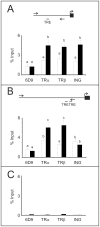
Figure 5. Chromatin immunoprecipitation assays indicate that ING protein associates with the TRβ and TH/bZIP promoters that are known to be T3-regulated in tadpole tails.
The promoter regions amplified upon ChIP are indicated by the arrows in the cartoons above the graphs for A) TRβ and B) TH/bZIP promoters and C) a β-actin gene control. The graphs show the percent input values of the amplicons obtained after immunoprecipitation with the indicated antibodies directed against TRα, TRβ, ING, and a control antibody, 6D9. DNA-protein complexes were obtained from pools of 7–12 tails from tadpoles NF stage 52–54 that were either time-matched solvent controls (white bars) or treated with 10 nM T3 for 48 h (black bars). Independent sets of animals were tested and averaged (n = 3–6, TRβ promoter; n = 3–5, TH/bZIP promoter). The error bars represent the standard error of the mean, and statistical significance (p<0.05, Mann Whitney U) is indicated by different letters.
Figure 6
Figure 6. Evidence that ING proteins are present in TR-containing complexes.
A) Endogenous p33ING protein coimmunoprecipitates with TRs in tadpole tail homogenates. Immunoprecipitations with a mouse polyclonal anti-TR antibody were carried out on X. laevis total tail homogenates from metamorphic climax NF stage 60–62 tadpoles (where ING, TH, and TR levels are all high), followed by immunoblots with a mouse monoclonal anti-ING antibody. A weak, but highly reproducible, specific 33 kDa band (indicated by an arrow) was detected (lane 2) when Sepharose beads were coincubated with anti-TR antibody and tail homogenate (Protein) and not present when either was incubated alone with the beads (lanes 3–4). Lane 1, protein homogenate (30 µg). This band is also not visible in Western blots where the anti-ING antibody was preincubated with either purified, bacterially-expressed p33ING1 (ING1 block; lanes 5–7) or p33ING2 (ING2 block; lanes 8–10). Lanes 11 and 12 are bacterially-expressed His-tagged Xenopus p33ING1 and p33ING2 proteins, respectively. Neither of the two slower migrating ING isoforms in lane 1 (see [49]) was immunoprecipitated. The strong upper and lower bands observed in lanes 2-10 are light and heavy chain immunoglobulins. B) Immunoblots of the same samples as above using another anti-TR antibody to demonstrate that TRs are immunoprecipitated. The 9B2 anti-TR antibody identified a band at ∼46 kDa that comigrates with bacterially-expressed frog TRα-His protein. The results shown represent one of three experiments with similar results.

Similar articles

References

    1. Menendez C, Abad M, Gomez-Cabello D, Moreno A, Palmero I. ING proteins in cellular senescence. Curr Drug Targets. 2009;10:406–417. - PubMed
    1. Unoki M, Kumamoto K, Takenoshita S, Harris CC. Reviewing the current classification of inhibitor of growth family proteins. Cancer Sci. 2009;100:1173–1179. - PMC - PubMed
    1. Li J, Wang Y, Wong RP, Li G. The role of ING tumor suppressors in UV stress response and melanoma progression. Curr Drug Targets. 2009;10:455–464. - PubMed
    1. Gunduz M, Demircan K, Gunduz E, Katase N, Tamamura R, Nagatsuka H. Potential usage of ING family members in cancer diagnostics and molecular therapy. Curr Drug Targets. 2009;10:465–476. - PubMed
    1. Walzak AA, Veldhoen N, Feng X, Riabowol K, Helbing CC. Expression profiles of mRNA transcript variants encoding the human inhibitor of growth tumor suppressor gene family in normal and neoplastic tissues. Exp Cell Res. 2008;314:273–285. - PubMed

Publication types

MeSH terms