Phages bearing affinity peptides to bovine rotavirus differentiate the virus from other viruses
- PMID: 22163050
- PMCID: PMC3232237
- DOI: 10.1371/journal.pone.0028667
Phages bearing affinity peptides to bovine rotavirus differentiate the virus from other viruses
Abstract
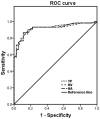
The aim of this study was to identify potential ligands and develop a novel diagnostic test to pathogenic bovine rotavirus (BRV) using phage display technology. The viruses were used as an immobilized target followed by incubation with a 12-mer phage display random peptide library. After five rounds of biopanning, phages had a specific binding activity to BRV were isolated. DNA sequencing indicated that phage displayed peptides HVHPPLRPHSDK, HATNHLPTPHNR or YPTHHAHTTPVR were potential ligands to BRV. Using the specific peptide-expressing phages, we developed a phage-based ELISA to differentiate BRV from other viruses. Compared with quantitative real-time PCR (qPCR), the phage-mediated ELISA was more suitable for the capture of BRV and the detection limitation of this approach was 0.1 µg/ml of samples. The high sensitivity, specificity and low cross-reactivity for the phage-based ELISA were confirmed in receiver operating characteristics (ROC) analysis.
Conflict of interest statement
Figures


Similar articles
-
Phages bearing affinity peptides to severe acute respiratory syndromes-associated coronavirus differentiate this virus from other viruses.J Clin Virol. 2013 Aug;57(4):305-10. doi: 10.1016/j.jcv.2013.04.002. Epub 2013 May 9. J Clin Virol. 2013. PMID: 23664850 Free PMC article.
-
Phage displayed peptides to avian H5N1 virus distinguished the virus from other viruses.PLoS One. 2011;6(8):e23058. doi: 10.1371/journal.pone.0023058. Epub 2011 Aug 22. PLoS One. 2011. PMID: 21887228 Free PMC article.
-
Highly sensitive detection of the group A Rotavirus using Apolipoprotein H-coated ELISA plates compared to quantitative real-time PCR.Virol J. 2011 Feb 10;8:63. doi: 10.1186/1743-422X-8-63. Virol J. 2011. PMID: 21310042 Free PMC article.
-
Phage display biopanning and isolation of target-unrelated peptides: in search of nonspecific binders hidden in a combinatorial library.Amino Acids. 2016 Dec;48(12):2699-2716. doi: 10.1007/s00726-016-2329-6. Epub 2016 Sep 20. Amino Acids. 2016. PMID: 27650972 Review.
-
Phage display: concept, innovations, applications and future.Biotechnol Adv. 2010 Nov-Dec;28(6):849-58. doi: 10.1016/j.biotechadv.2010.07.004. Epub 2010 Jul 23. Biotechnol Adv. 2010. PMID: 20659548 Review.
Cited by
-
Phage display for identifying peptides that bind the spike protein of transmissible gastroenteritis virus and possess diagnostic potential.Virus Genes. 2015 Aug;51(1):51-6. doi: 10.1007/s11262-015-1208-7. Epub 2015 May 27. Virus Genes. 2015. PMID: 26013256 Free PMC article.
-
Phages bearing affinity peptides to severe acute respiratory syndromes-associated coronavirus differentiate this virus from other viruses.J Clin Virol. 2013 Aug;57(4):305-10. doi: 10.1016/j.jcv.2013.04.002. Epub 2013 May 9. J Clin Virol. 2013. PMID: 23664850 Free PMC article.
-
Biopanning of polypeptides binding to bovine ephemeral fever virus G1 protein from phage display peptide library.BMC Vet Res. 2018 Jan 4;14(1):3. doi: 10.1186/s12917-017-1315-x. BMC Vet Res. 2018. PMID: 29301517 Free PMC article.
-
Altered specificity of single-chain antibody fragments bound to pandemic H1N1-2009 influenza virus after conversion of the phage-bound to the soluble form.BMC Res Notes. 2012 Sep 4;5:483. doi: 10.1186/1756-0500-5-483. BMC Res Notes. 2012. PMID: 22943792 Free PMC article.
-
Screening and antiviral analysis of phages that display peptides with an affinity to subunit C of porcine aminopeptidase.Monoclon Antib Immunodiagn Immunother. 2013 Oct;32(5):326-9. doi: 10.1089/mab.2013.0038. Monoclon Antib Immunodiagn Immunother. 2013. PMID: 24111863 Free PMC article.
References
-
- Estes MK, Kapikian AZ, In Knipe DM, Howley PM, Griffin DE, et al. 2001. pp. 1917–1974. Fields virology, 5th ed., vol.2. Lippincott Williams & Wilkins, Philadelphia, PA.
-
- Knipe DM, Howley PM, Griffin DE, Lamb RA, Martin MA, et al. Fields Virology, 4th ed., vol. 2. 2001;1747–1785 Lippincott, Williams and Wilkins, Philadelphia, Pennsylvania.
-
- Li X, Chang J, Liu H, Yu L. Seroepidemiological survey of the group A bovine rotavirus infection in China. Chines J Preventive Vet Med. 2010;9:664–667. (in Chinese).
Publication types
MeSH terms
Substances
LinkOut - more resources
Full Text Sources
Miscellaneous

