

Cannabidiol reduces Aβ-induced neuroinflammation and promotes hippocampal neurogenesis through PPARγ involvement
- PMID: 22163051
- PMCID: PMC3230631
- DOI: 10.1371/journal.pone.0028668
Cannabidiol reduces Aβ-induced neuroinflammation and promotes hippocampal neurogenesis through PPARγ involvement
Abstract
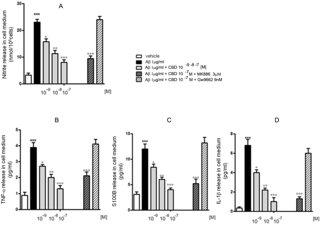
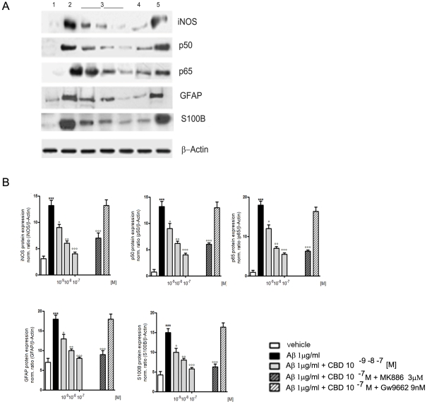
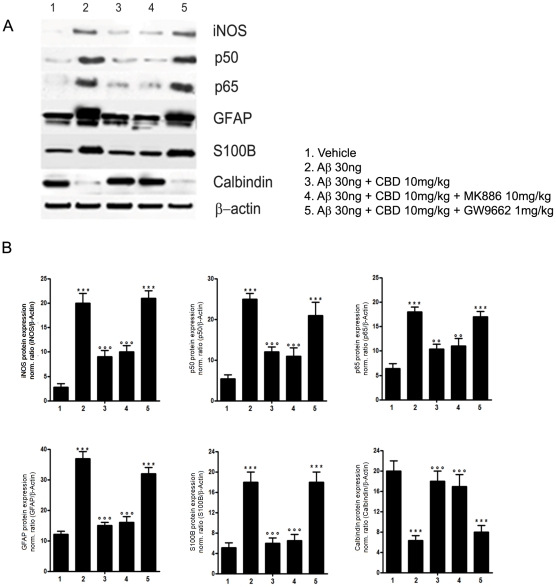
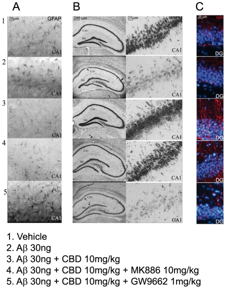
Peroxisome proliferator-activated receptor-γ (PPARγ) has been reported to be involved in the etiology of pathological features of Alzheimer's disease (AD). Cannabidiol (CBD), a Cannabis derivative devoid of psychomimetic effects, has attracted much attention because of its promising neuroprotective properties in rat AD models, even though the mechanism responsible for such actions remains unknown. This study was aimed at exploring whether CBD effects could be subordinate to its activity at PPARγ, which has been recently indicated as its putative binding site. CBD actions on β-amyloid-induced neurotoxicity in rat AD models, either in presence or absence of PPAR antagonists were investigated. Results showed that the blockade of PPARγ was able to significantly blunt CBD effects on reactive gliosis and subsequently on neuronal damage. Moreover, due to its interaction at PPARγ, CBD was observed to stimulate hippocampal neurogenesis. All these findings report the inescapable role of this receptor in mediating CBD actions, here reported.
Conflict of interest statement
Figures
Similar articles
-
Cannabidiol promotes amyloid precursor protein ubiquitination and reduction of beta amyloid expression in SHSY5YAPP+ cells through PPARγ involvement.Phytother Res. 2014 Jul;28(7):1007-13. doi: 10.1002/ptr.5095. Epub 2013 Nov 28. Phytother Res. 2014. PMID: 24288245
-
Effects of cannabidiol interactions with Wnt/β-catenin pathway and PPARγ on oxidative stress and neuroinflammation in Alzheimer's disease.Acta Biochim Biophys Sin (Shanghai). 2017 Oct 1;49(10):853-866. doi: 10.1093/abbs/gmx073. Acta Biochim Biophys Sin (Shanghai). 2017. PMID: 28981597
-
Cannabidiol in vivo blunts beta-amyloid induced neuroinflammation by suppressing IL-1beta and iNOS expression.Br J Pharmacol. 2007 Aug;151(8):1272-9. doi: 10.1038/sj.bjp.0707337. Epub 2007 Jun 25. Br J Pharmacol. 2007. PMID: 17592514 Free PMC article.
-
Cannabidiol goes nuclear: The role of PPARγ.Phytomedicine. 2023 Jun;114:154771. doi: 10.1016/j.phymed.2023.154771. Epub 2023 Mar 15. Phytomedicine. 2023. PMID: 36965374 Review.
-
Impact of Cannabis-Based Medicine on Alzheimer's Disease by Focusing on the Amyloid β-Modifications: A Systematic Study.CNS Neurol Disord Drug Targets. 2020;19(5):334-343. doi: 10.2174/1871527319666200708130745. CNS Neurol Disord Drug Targets. 2020. PMID: 32640965
Cited by
-
An Overview of Cannabidiol as a Multifunctional Drug: Pharmacokinetics and Cellular Effects.Molecules. 2024 Jan 18;29(2):473. doi: 10.3390/molecules29020473. Molecules. 2024. PMID: 38257386 Free PMC article. Review.
-
Molecular Targets of Cannabidiol in Neurological Disorders.Neurotherapeutics. 2015 Oct;12(4):699-730. doi: 10.1007/s13311-015-0377-3. Neurotherapeutics. 2015. PMID: 26264914 Free PMC article. Review.
-
Efficacy and Safety of the Ketogenic Diet for Mitochondrial Disease With Epilepsy: A Prospective, Open-labeled, Controlled Study.Front Neurol. 2022 Aug 1;13:880944. doi: 10.3389/fneur.2022.880944. eCollection 2022. Front Neurol. 2022. PMID: 35979062 Free PMC article.
-
From Cannabis sativa to Cannabidiol: Promising Therapeutic Candidate for the Treatment of Neurodegenerative Diseases.Front Pharmacol. 2020 Mar 6;11:124. doi: 10.3389/fphar.2020.00124. eCollection 2020. Front Pharmacol. 2020. PMID: 32210795 Free PMC article. Review.
-
Xanthoceraside modulates neurogenesis to ameliorate cognitive impairment in APP/PS1 transgenic mice.J Physiol Sci. 2018 Sep;68(5):555-565. doi: 10.1007/s12576-017-0561-9. Epub 2017 Jul 25. J Physiol Sci. 2018. PMID: 28744803 Free PMC article.
References
-
- Scuderi C, Filippis DD, Iuvone T, Blasio A, Steardo A, et al. Cannabidiol in medicine: a review of its therapeutic potential in CNS disorders. Phytother Res. 2009;23:597–602. - PubMed
-
- Iuvone T, Esposito G, Esposito R, Santamaria R, Di Rosa M, et al. Neuroprotective effect of cannabidiol, a non-psychoactive component from Cannabis sativa, on β-amyloid-induced toxicity in PC12 cells. J Neurochem. 2004;89:134–141. - PubMed
Publication types
MeSH terms
Substances
LinkOut - more resources
Full Text Sources
Medical
