Review
. 2010 Aug;1(2):289-305.
doi: 10.4155/tde.10.22.

Overcoming the challenges in the effective delivery of chemotherapies to CNS solid tumors

Affiliations
Review

Overcoming the challenges in the effective delivery of chemotherapies to CNS solid tumors

Hemant Sarin. Ther Deliv. 2010 Aug.

Abstract

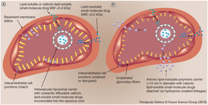
Locoregional therapies, such as surgery and intratumoral chemotherapy, do not effectively treat infiltrative primary CNS solid tumors and multifocal metastatic solid tumor disease of the CNS. It also remains a challenge to treat such CNS malignant solid tumor disease with systemic chemotherapies, although these lipid-soluble small-molecule drugs demonstrate potent cytotoxicity in vitro. Even in the setting of a 'normalized' tumor microenvironment, small-molecule drugs do not accumulate to effective concentrations in the vast majority of tumor cells, which is due to the fact that small-molecule drugs have short blood half-lives. It has been recently shown that drug-conjugated spherical lipid-insoluble nanoparticles within the 7-10 nm size range can deliver therapeutic concentrations of drug fraction directly into individual tumor cells following systemic administration, since these functionalized particles maintain peak blood concentrations for several hours and are smaller than the physiologic upper limit of pore size in the VEGF-derived blood capillaries of solid tumors, which is approximately 12 nm. In this article, the physiologic and ultrastructural basis of this novel translational approach for the treatment of CNS, as well as non-CNS, solid cancers is reviewed.


Conflict of interest statement

Financial & competing interests disclosure: The author has no other relevant affiliations or financial involvement with any organization or entity with a financial interest in or financial conflict with the subject matter or materials discussed in the manuscript apart from those disclosed.

Figures

Figure 1
Figure 1. Capillary wall ultrastructures of normal brain and spinal cord parenchyma blood capillaries and VEGF-derived solid tumor blood capillaries and transcapillary routes for passage of small molecules and drugs and macromolecular systemic chemotherapies
(A) Normal brain and spinal cord parenchyma blood capillary and (B) VEGF-derived solid tumor blood capillary. Capillary wall ultrastructures. Depicted are the three layers of the capillary wall. The yellow protrusions into the capillary lumen represent individual fibers of the polysaccharide-rich endothelial glycocalyx matrix with narrow interspacing. The bigger protrusions in (A) reflect the thicker glycocalyx of normal CNS tissue blood capillaries. The blue zona occludens interendothelial cell junctions between endothelial cells of normal brain and spinal cord parenchyma blood capillaries depict the intact functional interendothelial junctions of these blood capillaries, whereas the purple zona occludens interendothelial cell junctions of solid tumor tissue blood capillaries depict the disrupted dysfunctional interendothelial junctions of these blood capillaries. The small red domes represent the central membranous knobs of the diaphragmed fenestrae of the fenestrated endothelial cells of the VEGF-derived solid tumor blood capillaries. The pore openings of the diaphragmed fenestrae are wide enough to allow for the transcapillary passage of lipid-insoluble and cationic lipid-soluble molecules up to approximately 12 nm in diameter. The orange layer encircling the exterior of the endothelial cell lining layer depicts the collagenous lattice of the basement membrane layer. The basement membrane layers of both blood capillary types are depicted as being ultrastructurely similar, since the basement membranes of both blood capillary types are functionally intact. Transcapillary routes for passage of small molecules and drugs and macromolecular systemic chemotherapies. The small blue dots in the capillary lumens and the tissue interstitial spaces represent lipid-insoluble small molecules with molecular weights less than 0.2 kDa (e.g., electrolytes and nonelectrolytes), which can pass through the intact functional zona occludens interendothelial cell junctions of normal brain and spinal cord parenchyma blood capillaries and distribute into normal brain and spinal cord parenchyma interstitium during the time-period when the transcapillary concentration gradient is favoring forward diffusion from the capillary lumen into the tissue interstitium. The small green dots represent lipid-soluble molecule chemotherapy drugs with molecular weight less than 0.4 KDa (e.g., lomustine, 0.24 kDa), which can readily diffuse across the phospholipid bilayers of endothelial cell membranes and distribute nonselectively into normal healthy tissues, including normal brain and spinal cord parenchyma (shown), again, during the time-period when the transcapillary concentration gradient is favoring forward diffusion from the capillary lumen into the tissue interstitium. The small dots with a green exterior and blue interior represent charged cationic lipid-soluble small-molecule chemotherapy drugs of molecular weights greater than 0.4 kDa (e.g., doxorubicin, 0.54 kDa). Some are shown to be still encapsulated within the aqueous interior of a liposome, which is a large polymeric drug carrier for the purposes of sustained drug release, and remains within the capillary lumen, in the cases of both normal brain and spinal cord parenchyma and solid tumor blood capillaries. Those on the outside of the liposome represent the drug molecules that have diffused out of the liposome. Such free cationic lipid-soluble small drugs in the capillary lumen, although charged, cannot diffuse across the intact zona occludens interendothelial cell junctions of the capillary wall of normal brain and spinal cord parenchyma blood capillaries into normal brain and spinal cord parenchyma interstitium, which restrict the transcapillary passage of such charged drugs with molecular weights greater than 0.2 kDa. Such free cationic lipid-soluble small drugs in the capillary lumen, although lipid-soluble, also do not diffuse readily across the phospholipid bilayers of endothelial cell membranes, which restrict the transcapillary passage of such lipid-soluble small drugs with molecular weights greater than 0.4 kDa. The large structures containing multiple small dots represent anionic lipid-insoluble polymeric carriers less than 12 nm in diameter with cationic lipid-soluble small molecule drugs attached to the exterior via hydrazone covalent linkages (e.g., imageable dendrimer nanoparticles, with doxorubicin molecules covalently linked to surface groups via hydrolysable linkages, with diameters between 7 and 10 nm). Such lipid-insoluble polymeric drug carriers, with a neutralized exterior, upon systemic bolus administration, remain within the capillary lumens of normal brain and spinal cord parenchyma blood capillaries, but pass through the pore openings of the diaphragmed fenestrae of solid tumor blood capillaries and ‘selectively’ and ‘homogenously’ accumulate in the solid tumor tissue interstitium over time, delivering cytotoxic concentrations of chemotherapy drug into individual tumor cells.
Figure 2
Figure 2. T1-weighted dynamic contrast-enhanced MRI-based Gd concentration maps of Gd–dendrimer distribution over time within larger and smaller orthotopic RG-2 rodent malignant gliomas
(A) Larger RG-2 gliomas; The volume, in mm3, for each large orthotopic RG-2 glioma: Gd–G1, 104; Gd–G2, 94; Gd–G3, 94; LC Gd–G4, 162; standard Gd–G4, 200; Gd–G5, 230; Gd–G6, 201; Gd–G7, 170; and Gd–G8, 289. (B) Smaller RG-2 gliomas; The volume, in mm3, for each small orthotopic RG-2 glioma: Gd–G1, 27; Gd–G2, 28; Gd–G3, 19; LC Gd–G4, 24; standard Gd–G4, 17; Gd–G5, 18; Gd–G6, 22; Gd–G7, 24; and Gd–G8, 107. Respective Gd-dendrimer generations administered intravenously over 1 min at a Gd dose of 0.09 mmol Gd/kg animal body weight. Scale from 0 mM [Gd] to 0.1 mM [Gd]. G: Generation; LC: Lowly conjugated. Adapted with permission from [28].

Similar articles

Cited by

References

    1. Louis DN, Ohgaki H, Wiestler OD, et al. The 2007 WHO classification of tumours of the central nervous system. Acta Neuropathol. 2007;114(2):97. - PMC - PubMed
    1. Patchell RA, Tibbs PA, Walsh JW, et al. A randomized trial of surgery in the treatment of single metastases to the brain. N Engl J Med. 1990;322(8):494–500. - PubMed
    1. Weil RJ, Lonser RR, DeVroom HL, Wanebo JE, Oldfield EH. Surgical management of brainstem hemangioblastomas in patients with von Hippel-Lindau disease. J Neurosurg. 2003;98(1 Suppl):95–105. - PubMed
    1. Moss JM, Choi CY, Adler JR, Jr, Soltys SG, Gibbs IC, Chang SD. Stereotactic radiosurgical treatment of cranial and spinal hemangioblastomas. Neurosurgery. 2009;65(1):79–85. - PubMed
    1. Wohrer A, Waldhor T, Heinzl H, et al. The Austrian brain tumour registry: a cooperative way to establish a population-based brain tumour registry. J Neurooncol. 2009;95(3):401–411. - PubMed

Publication types

MeSH terms

LinkOut - more resources