. 2011;6(12):e27959.
doi: 10.1371/journal.pone.0027959. Epub 2011 Dec 7.

Redistribution of DAT/α-synuclein complexes visualized by "in situ" proximity ligation assay in transgenic mice modelling early Parkinson's disease

Affiliations

Redistribution of DAT/α-synuclein complexes visualized by "in situ" proximity ligation assay in transgenic mice modelling early Parkinson's disease

Arianna Bellucci et al. PLoS One. 2011.

Erratum in

  • PLoS One. 2012;7(1). doi:10.1371/annotation/58679d17-c619-4f13-b11f-146dacfe7e92. Grazia, Maria [corrected to Spillantini, Maria Grazia]

Abstract

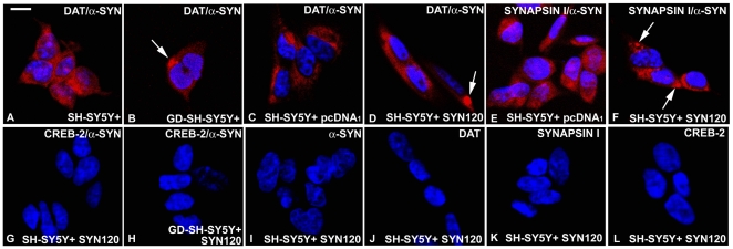
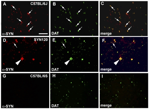
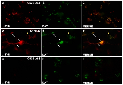
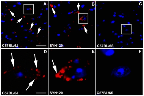
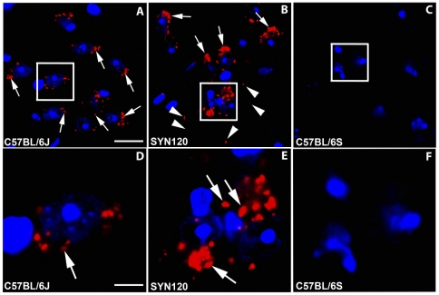
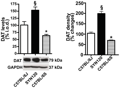
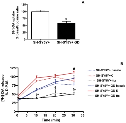
Alpha-synuclein, the major component of Lewy bodies, is thought to play a central role in the onset of synaptic dysfunctions in Parkinson's disease (PD). In particular, α-synuclein may affect dopaminergic neuron function as it interacts with a key protein modulating dopamine (DA) content at the synapse: the DA transporter (DAT). Indeed, recent evidence from our "in vitro" studies showed that α-synuclein aggregation decreases the expression and membrane trafficking of the DAT as the DAT is retained into α-synuclein-immunopositive inclusions. This notwithstanding, "in vivo" studies on PD animal models investigating whether DAT distribution is altered by the pathological overexpression and aggregation of α-synuclein are missing. By using the proximity ligation assay, a technique which allows the "in situ" visualization of protein-protein interactions, we studied the occurrence of alterations in the distribution of DAT/α-synuclein complexes in the SYN120 transgenic mouse model, showing insoluble α-synuclein aggregates into dopaminergic neurons of the nigrostriatal system, reduced striatal DA levels and an altered distribution of synaptic proteins in the striatum. We found that DAT/α-synuclein complexes were markedly redistributed in the striatum and substantia nigra of SYN120 mice. These alterations were accompanied by a significant increase of DAT striatal levels in transgenic animals when compared to wild type littermates. Our data indicate that, in the early pathogenesis of PD, α-synuclein acts as a fine modulator of the dopaminergic synapse by regulating the subcellular distribution of key proteins such as the DAT.


Conflict of interest statement

Competing Interests: The authors have declared that no competing interests exist.

Figures

Figure 1
Figure 1. Detection of DAT and α-synuclein interaction by PLA “in vitro.”
A: DAT and α-synuclein PLA in control SH-SY5Y+ cells. Please note a diffuse PLA-positive red signal. B: DAT and α-synuclein PLA in glucose deprived SH-SY5Y+ cells. Please note the presence of a PLA-positive inclusion (indicated by the arrow) within one of these cells. C: DAT and α-synuclein PLA in control SH-SY5Y+ cells. The PLA signal is weak and diffuse in these cells. D: DAT and α-synuclein PLA in SYN120-transfected SH-SY5Y+ cells. Please note the presence of an inclusion (indicated by the arrow) within one of these cells. E: synapsin I and α-synuclein PLA on pcDNA1-transfected SH-SY5Y cells. Please note that the PLA signal appears to be weak and diffused. F: synapsin I and α-synuclein PLA in SYN120-transfected SH-SY5Y+ cells. Some positive dots (indicated by the arrows) were visible within these cells. G: CREB-2 and α-synuclein PLA in control SH-SY5Y+ cells. No PLA signal was detected in these cells, which is indicative of the absence of CREB-2 and α-synuclein interaction. H: CREB-2 and α-synuclein PLA in glucose deprived-SYN120-transfected SH-SY5Y+ cells. No PLA signal is visible. I-L: Single labeled cells were used as negative controls for PLA experiments. I: α-synuclein antibody; J: DAT antibody; K: synapsin I antibody; L: CREB-2 antibody. Scale bar: 15 µm for A–L.
Figure 2
Figure 2. DAT and α-synuclein double staining in the striatum.
DAT (green signal) and α-synuclein (red signal) double staining in the striatum of C57BL/6J (panel A–C), SYN120 (panel D–F) and C57BL/6S (panel G–I) mice. Panels C, F, I show the merges of DAT and α-synuclein labeling. Please note the marked redistribution of both DAT and α-synuclein immunolabeling in the SYN120 mice. In the C57BL/6S mice which didn't express α-synuclein, DAT distribution was similar to that observed in the C57BL/6J mice. Scale bar: 60 µm for A–I.
Figure 3
Figure 3. DAT and α-synuclein double staining in the substantia nigra.
DAT (green signal) and α-synuclein (red signal) double staining in the substantia nigra of C57BL/6J (panel A–C), SYN120 (panel D–F) and C57BL/6S (panel G–I) mice. Panels C, F and I show the merges between DAT and α-synuclein immunolabelings. Please note that DAT and α-synuclein co-localized in dot-like clusters in the C57BL/6J mice. In the SYN120 mice DAT labeling was concentrated in big intracellular inclusions and within neuronal processes (arrows) and cell bodies (arrowhead) together with truncated α-synuclein, although some cells showed the same clustered co-localization of DAT and α-synuclein that we observed in the C57BL/6J mice (yellow arrow). In the C57BL/6S mice DAT distribution in the substantia was similar to that observed in the C57BL/6S mice. Scale bar: 40 µm for A–I.
Figure 4
Figure 4. DAT and α-synuclein PLA in the striatum.
DAT and α-synuclein PLA in the striatum of C57BL/6J (panel A) SYN120 (panel B) and C57BL/6S (panel C) mice. Panels D, E and F show higher magnifications of the squares in panels A, B and C, respectively. Please note that in the SYN120 mice the PLA signal (indicated by the arrows) displayed a more condensed distribution with respect to the C57BL/6J mice. In the C57BL/6S mice, which were lacking α-synuclein, no PLA signal was detected. Scale bars: A = 60 µm for A–C; D = 10 µm for D–F.
Figure 5
Figure 5. DAT and α-synuclein PLA signal in the substantia nigra.
DAT and α-synuclein PLA in the substantia nigra of C57BL/6J (panel A, D) SYN120 (panel B, E) and C57BL/6S (panel C, F) mice. Panels D, E and F show higher magnifications of the squares in panels A, B and C, respectively. In the C57BL/6J mice PLA-positive blobs were present while in SYN120 mice the PLA signal was condensed in big dots. In the C57BL/6S mice no PLA signal was detected. Scale bars: A = 60 µm for A–C; D = 20 µm for D–F.
Figure 6
Figure 6. DAT levels in the striatum.
A: Semiquantitative analysis of DAT levels in the striatum of C57BL/6J (white bars), SYN120 (black bars) and C57BL/6S (striped bars) mice. Please note the statistically significant increase of DAT levels in the SYN120 transgenic mice when compared to C57BL/6J and C57BL/6S wild type mice. B: Quantitative analysis of DAT immunoreactivity (% optical density) in the striatum of SYN120, C57BL/6J and C57BL/6S mice.
Figure 7
Figure 7. [3H]-DA uptake and [3H]-DA release in control and glucose-deprived SH-SY5Y+ cells.
A: The graph is showing [3H]-DA uptake by SH-SY5Y+ and glucose deprived SH-SY5Y+ cells (SH-SY5Y+ GD). B: The graph is showing [3H]-DA release by SH-SY5Y+ and SH-SY5Y+ GD cells in basal contition and after TTX block or 50 mM K+ (K) treatment.

References

    1. Spillantini MG, Schmidt ML, Lee VM, Trojanowski JQ, Jakes R, et al. Alpha-synuclein in Lewy bodies. Nature. 1997;388:839–840. - PubMed
    1. Cookson MR. The biochemistry of Parkinson's disease. Annu. Rev. Biochem. 2005;74:29–52. - PubMed
    1. Tofaris GK, Spillantini MG. Physiological and pathological properties of alpha-synuclein. Cell Mol. Life Sci. 2007;64:2194–2201. - PMC - PubMed
    1. Uversky VN. Neuropathology, biochemistry, and biophysics of alpha-synuclein aggregation. J Neurochem. 2007;103:17–37. - PubMed
    1. Kovacs GG, Milenkovic IJ, Preusser M, Budka H. Nigral burden of alpha-synuclein correlates with striatal dopamine deficit. Mov Disord. 2008;23:1608–1612. - PubMed

Publication types