Review
. 2011 Nov;3(11):2238-54.
doi: 10.3390/v3112238. Epub 2011 Nov 15.

Hepatitis C virus assembly imaging

Affiliations
Review

Hepatitis C virus assembly imaging

Costin-Ioan Popescu et al. Viruses. 2011 Nov.

Abstract

Hepatitis C Virus (HCV) assembly process is the least understood step in the virus life cycle. The functional data revealed by forward and reverse genetics indicated that both structural and non-structural proteins are involved in the assembly process. Using confocal and electron microscopy different groups determined the subcellular localization of different viral proteins and they identified the lipid droplets (LDs) as the potential viral assembly site. Here, we aim to review the mechanisms that govern the viral proteins recruitment to LDs and discuss the current model of HCV assembly process. Based on previous examples, this review will also discuss advanced imaging techniques as potential means to extend our present knowledge of HCV assembly process.

Keywords: cellular imaging; hepatitis C virus; virus assembly.


Figures

Figure 1.
Figure 1.
Representation of the genetic organization of hepatitis C virus (HCV). The N-terminal part of the genome comprises the structural elements core (C)—the forming unit of the capsid and the envelope glycoproteins, E1 and E2. The C-terminal part of the genome consists of the non-structural proteins (NS3 to NS5B) responsible for HCV genome amplification. p7 and NS2 are non-structural protein dispensable for replication, but essential for viral assembly. HCV proteins are synthesized as a polyprotein, which is processed by endogenous proteases (full circle—signal peptide peptidase, open diamond— signal peptidase) or viral encoded proteases: NS2 (full diamond) and NS3 (open circle) to generate 10 mature proteins.
Figure 2.
Figure 2.
Working model of HCV assembly. Viral assembly is triggered by the encounter of three modules: core, E1E2p7NS2 complex and the replication complex (RC). The assembly site is supposed to be in the membranous microenvironment of the lipid droplet (LD) and the endoplasmic reticulum (ER). The driving force of viral budding potentially comes from three directions: pushing force of the nascent nucleocaspsid, the pulling force of envelope proteins which might stabilize the viral surface architecture by intermolecular disulfide bridges and the force of the nascent luminal LD (luLD) between the ER leaflets. The result is a hybrid lipoviroparticle, which acquires ApoE presumably by its lipid component and in primary hepatocytes the lipoprotein moiety may mature into a VLDL-like structure (adapted from [46]). MTP stands for microsomal triglyceride transfer protein that is involved in the VLDL pathway. sER/mw is for smooth ER/ membranous web where the HCV replication is believed to take place.
Figure 3.
Figure 3.
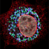
NS2 protein accumulates at the site of virus assembly. Confocal image of an electroporated hepatocyte with the HCV genome shows the accumulation of NS2 (red) and core protein (blue) in close proximity to cytoplasmic LDs (green), the putative site of HCV assembly. The nucleus was counterstained with DAPI (grey).

Similar articles

Cited by

References

    1. Shepard C.W., Finelli L., Alter M.J. Global epidemiology of hepatitis C virus infection. Lancet Infect. Dis. 2005;5:558–567. - PubMed
    1. Lindenbach B.D., Thiel H.J., Rice C.M. Flaviviridae: The viruses and their replication. In: Knipe D.M., Howley P.M., editors. Fields Virology. 5th ed. Lippincott Williams & Wilkins; Philadelphia, PA, USA: 2007. pp. 1101–1152.
    1. Lohmann V., Korner F., Koch J., Herian U., Theilmann L., Bartenschlager R. Replication of subgenomic hepatitis C virus RNAs in a hepatoma cell line. Science. 1999;285:110–113. - PubMed
    1. Bartosch B., Dubuisson J., Cosset F.L. Infectious hepatitis C pseudo-particles containing functional e1e2 envelope protein complexes. J. Exp. Med. 2003;197:633–642. - PMC - PubMed
    1. Drummer H.E., Maerz A., Poumbourios P. Cell surface expression of functional hepatitis C virus e1 and e2 glycoproteins. FEBS Lett. 2003;546:385–390. - PubMed

Publication types