

Hepatitis C virus assembly imaging
- PMID: 22163343
- PMCID: PMC3230850
- DOI: 10.3390/v3112238
Hepatitis C virus assembly imaging
Abstract
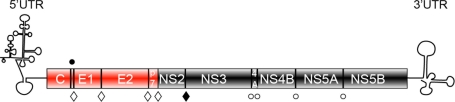
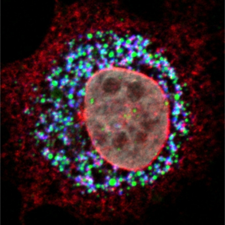
Hepatitis C Virus (HCV) assembly process is the least understood step in the virus life cycle. The functional data revealed by forward and reverse genetics indicated that both structural and non-structural proteins are involved in the assembly process. Using confocal and electron microscopy different groups determined the subcellular localization of different viral proteins and they identified the lipid droplets (LDs) as the potential viral assembly site. Here, we aim to review the mechanisms that govern the viral proteins recruitment to LDs and discuss the current model of HCV assembly process. Based on previous examples, this review will also discuss advanced imaging techniques as potential means to extend our present knowledge of HCV assembly process.
Keywords: cellular imaging; hepatitis C virus; virus assembly.
Figures

References
-
- Shepard C.W., Finelli L., Alter M.J. Global epidemiology of hepatitis C virus infection. Lancet Infect. Dis. 2005;5:558–567. - PubMed
-
- Lindenbach B.D., Thiel H.J., Rice C.M. Flaviviridae: The viruses and their replication. In: Knipe D.M., Howley P.M., editors. Fields Virology. 5th ed. Lippincott Williams & Wilkins; Philadelphia, PA, USA: 2007. pp. 1101–1152.
-
- Lohmann V., Korner F., Koch J., Herian U., Theilmann L., Bartenschlager R. Replication of subgenomic hepatitis C virus RNAs in a hepatoma cell line. Science. 1999;285:110–113. - PubMed
-
- Drummer H.E., Maerz A., Poumbourios P. Cell surface expression of functional hepatitis C virus e1 and e2 glycoproteins. FEBS Lett. 2003;546:385–390. - PubMed
Publication types
MeSH terms
Substances
LinkOut - more resources
Full Text Sources
Medical
