Review
. 2012:2012:519238.
doi: 10.1155/2012/519238. Epub 2011 Oct 25.

Ultrasound biomicroscopy in small animal research: applications in molecular and preclinical imaging

Affiliations
Review

Ultrasound biomicroscopy in small animal research: applications in molecular and preclinical imaging

A Greco et al. J Biomed Biotechnol. 2012.

Abstract

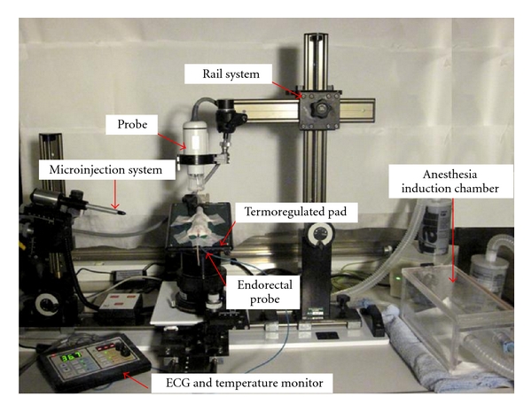
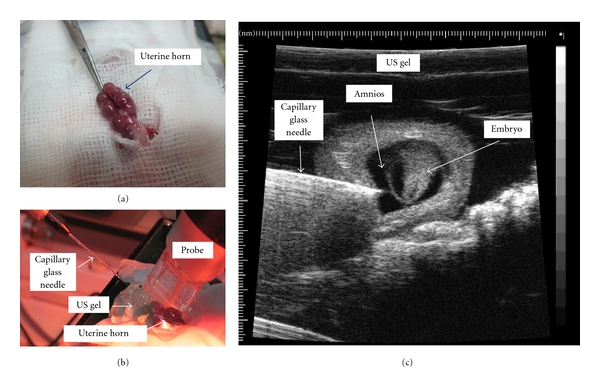
Ultrasound biomicroscopy (UBM) is a noninvasive multimodality technique that allows high-resolution imaging in mice. It is affordable, widely available, and portable. When it is coupled to Doppler ultrasound with color and power Doppler, it can be used to quantify blood flow and to image microcirculation as well as the response of tumor blood supply to cancer therapy. Target contrast ultrasound combines ultrasound with novel molecular targeted contrast agent to assess biological processes at molecular level. UBM is useful to investigate the growth and differentiation of tumors as well as to detect early molecular expression of cancer-related biomarkers in vivo and to monitor the effects of cancer therapies. It can be also used to visualize the embryological development of mice in uterus or to examine their cardiovascular development. The availability of real-time imaging of mice anatomy allows performing aspiration procedures under ultrasound guidance as well as the microinjection of cells, viruses, or other agents into precise locations. This paper will describe some basic principles of high-resolution imaging equipment, and the most important applications in molecular and preclinical imaging in small animal research.


Figures

Figure 1
Figure 1
Ultrasound biomicroscopy integrated mechanical support with rail system. The microinjection system for visualization and guidance of injection and extraction procedures in real time. Ultrasound probe is mounted on a system securing the probe in a stationary position when the ultrasound scan is in the desired image plane. After anesthesia, the mouse is positioned on a termoregulated pad to monitor ECG and temperature (with endorectal probe) and ensure mouse comfort during imaging.
Figure 2
Figure 2
Various view of three-dimensional reconstructions of a subcutaneous tumor from two bidimensional successive parallel cross-section images. (a) Cube view. On the bottom, the segmentational volumetric analysis with a volume estimation of 48.02 mm3. (b) Surface view. (c) Cross view. (d) Quantitative analysis of tumor growth in a mice model of mammary tumor. The tumor growth curve over time is showed in the diagram.
Figure 3
Figure 3
Behavior of microbubbles depends on the amplitude of ultrasound to which they are exposed. At very low acoustic power (mechanical index <0.05–0.1), microbubble oscillates in relatively symmetrical backscattering at the same frequency of incident ultrasound. At a slightly higher mechanical index of 0.1–0.3, the microbubble becomes somewhat oscillates in a nonlinear manner (nonlinear response), backscattering a variety of frequencies (harmonic). Higher acoustic pressures (MI > 0.3–0.6) destroy the microbubbles with high-intensity backscatter response.
Figure 4
Figure 4
Schematic representation of active targeting of biotin-streptavidine bridges. Streptavidine is used for attachment of biotinylated ligands onto the shell of ultrasound contrast microbubbles. Molecularly targeted microbubbles selective bind to sites of molecular expression on the endothelium.
Figure 5
Figure 5
Targeted US of endothelial antigens in vessels of a tumor tissue. On the top of the figure time/video intensity analysis before and after high-power destructive pulse and bottom a diagram representation of destructive methodology. Endothelial cells of vessels (orange) of tumor tissues expresses specific antigens. After intravenous administration targeted microbubbles float in vessels and remaining exclusively in the vascular compartment. Many of them bind to antigens of endothelial cells, whereas others remains in the vessel lumen freely circulating. After high-power destructive pulse, all microbubbles are destroyed (bound + circulating), following circulating microbubbles that arrive from outside of scan plane, which remain freely circulating for several seconds. Contrast intensity is the sum of the intensity from tissue, intensity from microbubbles not bound to receptors (circulating microbubbles), and intensity from microbubbles bound to receptors on endothelial cells. After digital subtraction of the video intensity calculated on 300 predestruction frames from video intensity calculated on 300 postdestruction frames, resulting video intensity is due only to bound microbubbles.
Figure 6
Figure 6
Cannulation of jugular and tail veins. The anesthetized mouse is immobilized by taping its harms on a termoregulated pad. A side of the neck or the tail of the mouse are disinfected. A 27-gauge needle connected by polyethylene tubing (PE10) to a 1 mL saline-filled cannula is inserted into the jugular vein or in the lateral tail vein approximately 2 cm from the body. The cannula, filled with heparin solution (750 units/mL in water), is inserted into the vein in caudal direction and fixed to the vein with cyanoacrylate glue (Histoacryl, B Braun, Am Aesculap-Platz, Germany). The proof of the correct position of the needle is given by blood visualization in the cannula. (a) Cannulation of jugular vein. (b) Cannulation of lateral tail vein.
Figure 7
Figure 7
In utero microinjection: laparoscopy along the linea alba. The uterine horns are gently exteriorized to record implantation sites and simply positioned on sterile gauze and covered with sterile acoustic gel (a). A 40 Mhz probe is used to image in real time the embryos, and the microinjection is performed by an automatic microinjector equipped with a capillary glass needle. The needle is advanced with the use a micromanipulator under echo guidance until the needle tip is in the desired location (b). UBM images of a capillary glass needle advanced under echo guidance until the needle tip is in the amnions (c).
Figure 8
Figure 8
(a) UBM microimaging of thyroid in living mice performed with a 35 MHz probe in a genetically engineered mice model of thyroid disease (TRK). The figure shows an enlarged left lobe with hypoechoic nodule (arrows). (b) Histopathology analysis shows a thyroid adenoma. From Mancini et al. Endocrinology [2].
Figure 9
Figure 9
Multimodality imaging of the lesion: human mammary tumor cells were implanted in the mammary fat pad of an athymic nude mice, 6 weeks old. There is a good correspondence between the morphology and the size of the tumor detected by UBM and necroscopic findings. (a) Bioluminescence of the tumor. (b) 18F-flourothymidine total body PET, tumor uptake is evident (arrow). (c) UBM with measurements of neoplasia. (d) Post mortem dimension of the explanted tumor.
Figure 10
Figure 10
(a) E9.5: heart tube and doppler spectral trace of ventricular inflow and outflow at 9.5 gestational age. (b) E11.5: doppler spectral trace of ascending aorta outflow at 11.5 gestational age. (c) E12.5: bidimensional image of atrio-ventricular chambers (a–V) interventricular septum (IVS); and doppler spectral trace of ascending aorta outflow at 12.5 gestational age. (d) E13.5: bidimensional image of atrioventricular chambers, interventricular septum, and left ventricle wall (LV wall); doppler spectral trace of ventricular inflow and outflow at 13.5 gestational age. The atrial wave (A-wave) was dominant. The rapid ventricular filling (E-wave) is a measure of ventricular compliance. (e) E15.5: bidimensional image of ventricular chambers, interventricular septum, and left ventricle wall in telesystolic and telediastolic fase of heart cycle in a 15.5 days mouse embryo.

References

    1. Hinton RB, Alfieri CM, Witt SA, et al. Mouse heart valve structure and function: echocardiographic and morphometric analyses from the fetus through the aged adult. American Journal of Physiology—Heart and Circulatory Physiology. 2008;294(6):H2480–H2488. - PubMed
    1. Mancini M, Vergara E, Salvatore G, et al. Morphological ultrasound microimaging of thyroid in living mice. Endocrinology. 2009;150(10):4810–4815. - PubMed
    1. Shen ZP, Brayman AA, Chen L, Miao CH. Ultrasound with microbubbles enhances gene expression of plasmid DNA in the liver via intraportal delivery. Gene Therapy. 2008;15(16):1147–1155. - PMC - PubMed
    1. Foster FS, Zhang M, Duckett AS, Cucevic V, Pavlin CJ. In vivo imaging of embryonic development in the mouse eye by ultrasound biomicroscopy. Investigative Ophthalmology and Visual Science. 2003;44(6):2361–2366. - PubMed
    1. Spurney CF, Lo CW, Leatherbury L. Fetal mouse imaging using echocardiography: a review of current technology. Echocardiography. 2006;23(10):891–899. - PubMed

LinkOut - more resources