Review
. 2010;10(12):11352-68.
doi: 10.3390/s101211352. Epub 2010 Dec 13.

Sensing-applications of surface-based single vesicle arrays

Affiliations
Review

Sensing-applications of surface-based single vesicle arrays

Sune M Christensen et al. Sensors (Basel). 2010.

Abstract

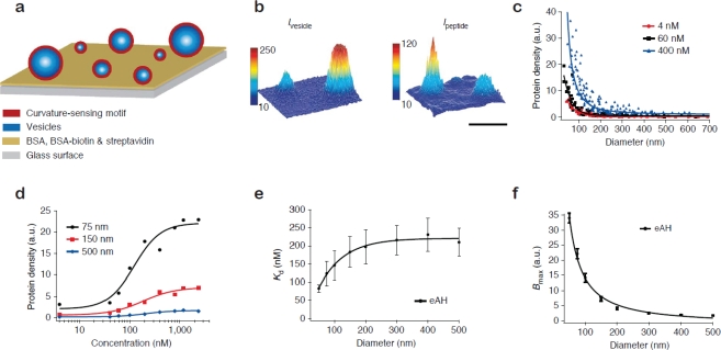
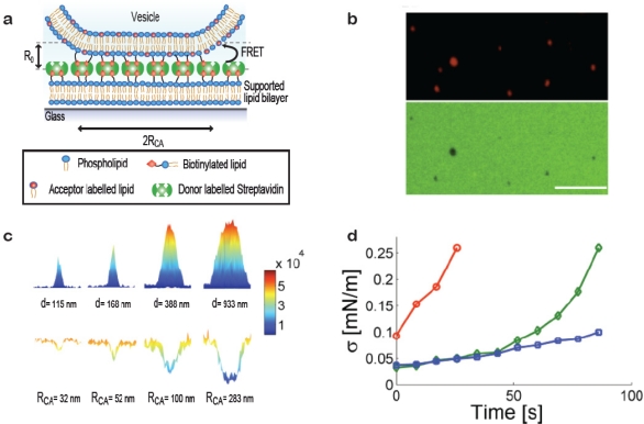
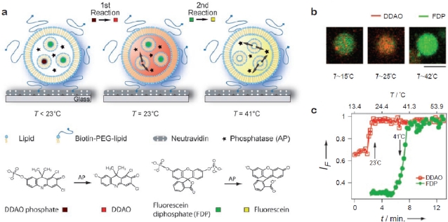
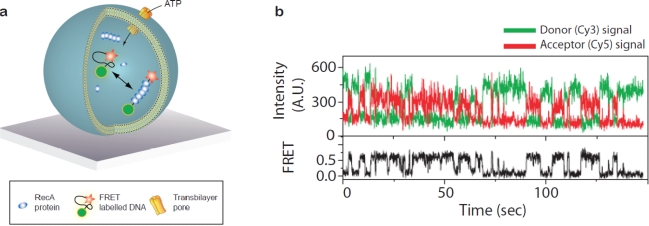
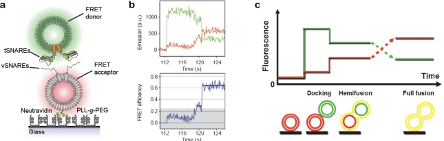
A single lipid vesicle can be regarded as an autonomous ultra-miniaturised 3D biomimetic "scaffold" (Ø≥13 nm) ideally suited for reconstitution and interrogation of biochemical processes. The enclosing lipid bilayer membrane of a vesicle can be applied for studying binding (protein/lipid or receptor/ligand interactions) or transmembrane events (membrane permeability or ion channel activation) while the aqueous vesicle lumen can be used for confining few or single macromolecules and probe, e.g., protein folding, catalytic pathways of enzymes or more complex biochemical reactions, such as signal transduction cascades. Immobilisation (arraying) of single vesicles on a solid support is an extremely useful technique that allows detailed characterisation of vesicle preparations using surface sensitive techniques, in particular fluorescence microscopy. Surface-based single vesicle arrays allow a plethora of prototypic sensing applications in a high throughput format with high spatial and high temporal resolution. In this review we present a series of applications of single vesicle arrays for screening/sensing of: membrane curvature dependent protein-lipid interactions, bilayer tension, reactions triggered in the vesicle lumen, the activity of transmembrane protein channels and biological membrane fusion reactions.

Keywords: model membrane systems; nanoreactors; single vesicles; vesicles.


Figures

Figure 1.
Figure 1.
Surface-based lipid vesicle systems. (a) Sketch showing examples on various biochemical processes reconstituted in an immobilised vesicle. (b) Surface plot of the fluorescence intensity of a vesicle stained with a membrane anchored dye. Quantification of the fluorescence signal of single vesicles provides access to information on vesicle size and allows following reactions taking place at the membrane or within the lumen. (c) Single vesicle array interrogated by fluorescence microscopy. (d) Fluorescence micrograph of a single vesicle array functionalised with two populations of vesicles (red and green labels, respectively) [1]. Bars: 10 μm and 0.5 μm. d was adapted from reference [1] (Copyright Wiley-VCH Verlag GmbH & Co. KGaA. Reproduced with permission).
Figure 2.
Figure 2.
Screening of membrane curvature dependent protein-lipid interactions. (a) Vesicles of different size, and therefore different membrane curvature, were immobilised on a glass coverslip and incubated with a curvature-sensing motif. (b) Single vesicle positions and sizes (curvatures) were assessed from the fluorescence intensity of a lipophilic dye incorporated in the membrane (left). The amount of bound fluorescently labelled protein (in the shown example the amphipathic helix containing N-terminal domain of endophilin A1, eAH) was recorded in a second channel. The surface plots of intensity clearly demonstrates curvature specific binding, i.e., the small vesicle to the left accommodated much higher density of protein than the large vesicle to the right. Bar: 1 μm. (c) Density of bound protein as a function of vesicle diameter for three different protein concentrations. (d) Binding curves as a function of vesicle diameter. (e,f) Kd and Bmax as a function of vesicle diameter extracted by fitting of binding curves as illustrated in (d). We acknowledge Nature Chemical Biology where the material in this figure originally appeared [17].
Figure 3.
Figure 3.
Measuring membrane tension on single vesicles by quantification of nanoscale contact areas formed between vesicles and a surface. (a) Sketch of the contact area formed between a vesicle and a streptavidin decorated supported bilayer. Quantitative information on the radius of the contact area (RCA) was obtained by measuring FRET between donor dyes on the streptavidin layer and acceptor dyes in the vesicle membrane. R0 depicts the Förster radius. (b) Fluorescence micrographs of vesicles with radius ≈100 nm (top) and the bilayer (bottom). FRET footprints are observed as a reduction in donor intensity. Bar: 5 μm. (c) Surface plots of fluorescence intensity of immobilised vesicles (top) and corresponding plots of fluorescence from the donor labelled streptavidin layer (bottom). By comparing theoretical and experimental results the degree of vesicle deformation upon immobilisation was evaluated. The obtained vesicle diameters, d, and the radii of the mapped contact areas (RCA) are indicated below the single vesicle intensity plots. (d) Time-resolved measurements of single vesicle contact areas used to evaluate the tension in the bilayer as a function of time. In this example tension increased due to strong laser illumination [22]. The graph shows data for 3 vesicles of different size (circles; vesicle diameter d = 362 nm, diamonds; d = 778 nm and squares; d = 1,272 nm). Figure adapted from [19].
Figure 4.
Figure 4.
Self-assembled nanoscale fluidic device; consecutive enzymatic reactions triggered in single giant unilamellar vesicles. (a) A giant vesicle immobilised on a neutravidin-coated glass coverslip. Alkaline phosphatase (stars) was incorporated in the giant vesicle together with two different sets of small unilamellar vesicles (SUVs) each loaded with a different non-fluorescent substrate for the enzyme. One set of SUVs (Tt ≈ 23 °C) contained dichlorodimethylacridinone (DDAO) phosphate (dark red) and the second SUV population (Tt ≈ 41 °C) carried fluorescein diphosphate (FDP, dark green). An increase of temperature triggered the release of the substrates in two consecutive steps at the corresponding phase transition temperatures. After release from the SUVs, the substrates were confined in the lumen of the giant vesicle where they were processed to their respective fluorescent products by the enzyme. (b) Fluorescence micrographs of the process depicted in a. Bar: 10 μm. (c) Fluorescence intensity traces demonstrating sequential release of substrates and their conversion to fluorescent products in a single giant vesicle during a temperature ramp. Figure adapted from [32] (Copyright Wiley-VCH Verlag GmbH & Co. KGaA. Reproduced with permission).
Figure 5.
Figure 5.
Single molecule recording of protein-DNA interactions driven by ATP inside a single vesicle. (a) A single strand of DNA labelled with a FRET pair encapsulated inside a surface-tethered vesicle together with the ATPase RecA (approximately seven copies per vesicle). In the cell RecA is active in DNA repair upon ATP regulated polymerisation on DNA. Upon RecA assembly the DNA was stretched giving rise to a reduction in FRET. Vesicles were made permeable to small molecules (<1 nm diameter) via pores in the membrane introduced either by utilising a lipid mixture with a phase transition at the temperature of the experiment [49] or using he bacterial toxin alpha-hemolysin [50]. Repeated polymerisation/dissociation reactions were triggered by exchange of ATP through the pores. (b) Fluctuations between stretched (low FRET) and unstretched (high FRET) states of the DNA. Figure b adapted from [49], copyright 2007 National Academy of Sciences, USA.
Figure 6.
Figure 6.
Single vesicle-vesicle fusion assay. (a) A population of vesicles carrying reconstituted vSNAREs is tethered to a solid support through biotin-neutravidin coupling and allowed to interact with a freely diffusing population of vesicles harbouring cognate tSNAREs. The surface is passivated with PLL-g-PEG to prevent non-specific interactions of the tSNARE vesicles and the surface. (b) Discrete steps of docking, hemifusion and full fusion as evidenced from the apparent FRET efficiency (bottom) calculated from donor (green) and acceptor (red) emission during fusion of a single vesicle pair. The abrupt increase in donor emission in the beginning of the trace marks vesicle docking. (c) Simplified sketch of the evolution of donor and acceptor emission and the configurations thought to be associated with each level of FRET. Figures (b,c) were adapted from [56], Copyright 2006 National Academy of Sciences, USA.

Similar articles

Cited by

References

    1. Stamou D, Duschl C, Delamarche E, Vogel H. Self-Assembled Microarrays of Attoliter Molecular Vessels. Angew Chem Int Ed. 2003;42:5580–5583. - PubMed
    1. Szoka F, Papahadjopoulos D. Comparative Properties and Methods of Preparation of Lipid Vesicles (Liposomes) Annu Rev Biophys Bioeng. 1980;9:467–508. - PubMed
    1. Edwards KA, Baeumner AJ. Analysis of Liposomes. Talanta. 2006;68:1432–1441. - PubMed
    1. Christensen SM, Stamou D. Surface-Based Lipid Vesicle Reactor Systems: Fabrication and Applications. Soft Matter. 2007;3:828–836. - PubMed
    1. Li J, Möhwald H, Ana Z, Lua G. Molecular Assembly of Biomimetic Microcapsules. Soft Matter. 2005;1:259–264. - PubMed

Publication types

MeSH terms

LinkOut - more resources