Review
. 2010;10(7):6623-61.
doi: 10.3390/s100706623. Epub 2010 Jul 9.

Microfluidic systems for biosensing

Affiliations
Review

Microfluidic systems for biosensing

Kuo-Kang Liu et al. Sensors (Basel). 2010.

Abstract

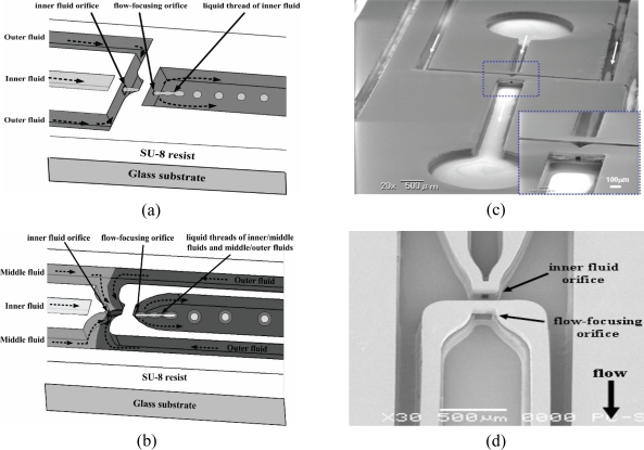
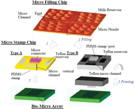
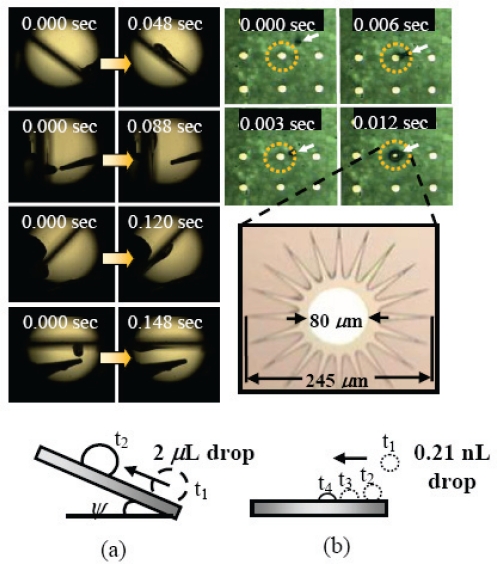
In the past two decades, Micro Fluidic Systems (MFS) have emerged as a powerful tool for biosensing, particularly in enriching and purifying molecules and cells in biological samples. Compared with conventional sensing techniques, distinctive advantages of using MFS for biomedicine include ultra-high sensitivity, higher throughput, in-situ monitoring and lower cost. This review aims to summarize the recent advancements in two major types of micro fluidic systems, continuous and discrete MFS, as well as their biomedical applications. The state-of-the-art of active and passive mechanisms of fluid manipulation for mixing, separation, purification and concentration will also be elaborated. Future trends of using MFS in detection at molecular or cellular level, especially in stem cell therapy, tissue engineering and regenerative medicine, are also prospected.

Keywords: MEMS; Micro total analysis systems (μTAS); droplet-based; drug delivery; lab-on-a-chip; microfluidic; stem cell; tissue engineering.


Figures

Figure 1.
Figure 1.
Schematic of one idealized total analysis device showing the various functions on a micro fluidic chip [8].
Figure 2.
Figure 2.
(a) Photographic overhead view of the μCEC chip (dark area in the central channel consisting of MWCNTs array, 1: sample reservoir, 2: eluent buffer/running buffer reservoir, 3: sample waste, 4: eluent buffer waste, 5: running buffer waste, D: detector), (b) The schematic arrangement of MWCNTs in μCEC channels as vertically aligned nanopillars, (c) the SEM image of a cross section (a-a’ plane) of the μCEC channel, (d) The SEM image of vertically-aligned MWCNTs directly grown in microchannel [65].
Figure 3.
Figure 3.
The schematics of (a) flow field evolution for DAEKF in a capillary electrophoresis nanochannel (solid black arrows represent analyte flow direction, and ζ2>ζ1>ζ3>ζ0), (b) detailed flow fields of five different regions for the DAEKF system in a nanochannel. (Region I: a 2-D shear flow, Region II: pulling effect-asymmetric electroosmotic flow (AEOF), Regions IV: pushing effect AEOF). Fluorescence image of Rhodamine B for (c) traditional EOF in a nanochannel, (d) the restacking effect by the DAEKF system in a nanochannel [66].
Figure 3.
Figure 3.
The schematics of (a) flow field evolution for DAEKF in a capillary electrophoresis nanochannel (solid black arrows represent analyte flow direction, and ζ2>ζ1>ζ3>ζ0), (b) detailed flow fields of five different regions for the DAEKF system in a nanochannel. (Region I: a 2-D shear flow, Region II: pulling effect-asymmetric electroosmotic flow (AEOF), Regions IV: pushing effect AEOF). Fluorescence image of Rhodamine B for (c) traditional EOF in a nanochannel, (d) the restacking effect by the DAEKF system in a nanochannel [66].
Figure 4.
Figure 4.
Schematic diagrams of the MEMS-based 3D MFFD using three layers of SU-8 resist for formation of (a) single emulsions and (b) double emulsions. (c) SEM images of the 3D MFFD for formation of single emulsions with inset showing the flow-focusing orifice with dimensions of 50 μm(W) × 50 μm(H). (d) SEM image of the 3D MFFD configuration for formation of double emulsions. The inner fluid orifice measuring 100 μm(W) × 50μm(H) × 100 μm(L) and the flow-focusing orifice measuring 200 μm(W) × 50 μm(H) × 100 μm(L) are coaxial, separated by a distance of 200 μm. The total height of the microchannels is 250 μm. (reprinted with permission from ref. , Copyright 2006, IOP).
Figure 5.
Figure 5.
Schematic of microarray system for batch-filling and in parallel printing of multiple proteins. (1) The micro connectors of the micro stamp chips are connected into the nozzles of the micro filling chip, and then the bio-fluids are transferred into the micro stamp simultaneously. (2) After the filling has been completed, those two chips are separated, and then PDMS stamps are used to print in parallel numerous arrays (reprinted with permission from ref. , ©2008 IEEE).
Figure 6.
Figure 6.
(a) Side view images of a 2 μL surface-ascending water droplet under different inclination angles θ (40°, 90°, 130°, and 180°) moving along the gradient with ψ = 8° and L = 12 mm. (b) A time sequence of top view images of a self-directed subnanoliter water droplet (0.21 nL) moving on a device with an array of circulating wedge-shape gradients. The dashed line indicates the coverage area of the gradients with the detailed dimensions shown in the enlarged view (after photoresist development) of the device. The arrows indicate the position of the droplet of which is transient (reprinted with permission from ref. , Copyright 2009, American Institute of Physics).
Figure 7.
Figure 7.
A continuous-flow PCR device where microfluidic droplets move through a temperature gradient toward the radial direction. The device contains an oil inlet (A) that joins an aqueous inlet channels (B) to form droplets (C). The droplets pass through the inner circles in the hot zone (D) to ensure initial denaturation of the template and travel on to the periphery where primer annealing and template extension occur (E). The droplets then flow back to the centre, where the DNA is denatured and a new cycle begins. For illustration, only 7 cycles are demonstrated, but 34 cycles have been achieved in [116].
Figure 8.
Figure 8.
Schematic diagram of MFFD for production of copolymer particles via in-situ UV polymerization by co-flow of aqueous (A) and comonomer (B) phases. Typical fluorescent images of the copolymer particles conjugated with IgG-Cy3 for CAA = 40 wt%. Fluid A: DI water + 2 wt% SDS, Fluid B: monomer (ethyleneglycol dimethacrylate, EGDMA) + 0∼40 wt% acrylic acid (AA) + 4wt% photoinitiator (HCPK, 1-hydroxycyclohexyl phenyl ketone) [140].
Figure 9.
Figure 9.
The scheme of the microfluidic device for the fabrication of photonic crystal beads by means of evaporation or UV polymerization to aggregate the colloidal nanoparticles that self-assembled in close-packed structures [156].

References

    1. Squires TM, Quake SR. Microfluidics: fluid physics at the nanoliter scale. Rev. Mod. Phys. 2005;77:977–1026.
    1. Bringer MR, Gerdts CJ, Song H, Tice JD, Ismagilov RF. Microfluidic systems for chemical kinetics that rely on chaotic mixing in droplets. Phil. Trans. R. Soc. Lond. A. 2004;362:1087–1104. - PMC - PubMed
    1. Weibel DB, Whitesides GM. Applications of microfluidics in chemical biology. Curr. Opin. Chem. Biol. 2006;10:584–591. - PubMed
    1. Breslauer DN, Lee PJ, Lee LP. Microfluidics-based system biology. Mol. Biosyst. 2006;2:97–112. - PubMed
    1. Hong J, Edel JB, deMello AJ. Micro-and nanofluidic systems for high-throughput biological screening. Drug Discovery Today. 2009;14:134–146. - PubMed

LinkOut - more resources