

A nanosensor for TNT detection based on molecularly imprinted polymers and surface enhanced Raman scattering
- PMID: 22163761
- PMCID: PMC3231613
- DOI: 10.3390/s110302700
A nanosensor for TNT detection based on molecularly imprinted polymers and surface enhanced Raman scattering
Abstract
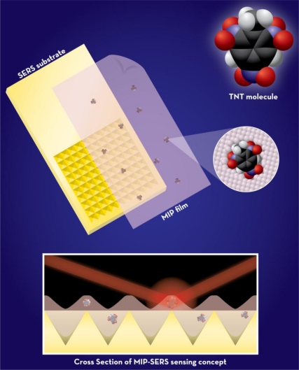
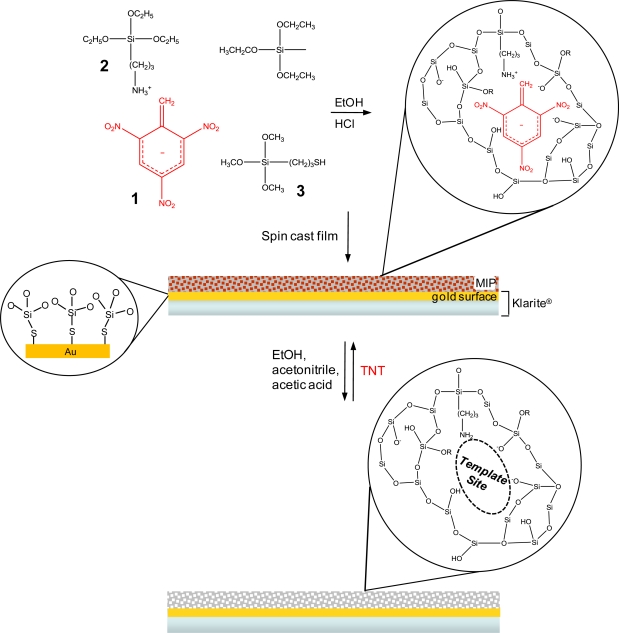
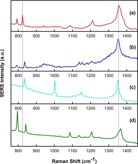
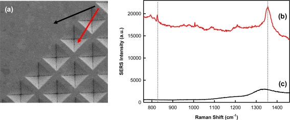
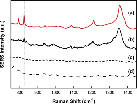
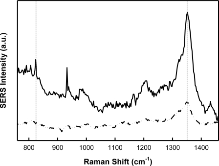
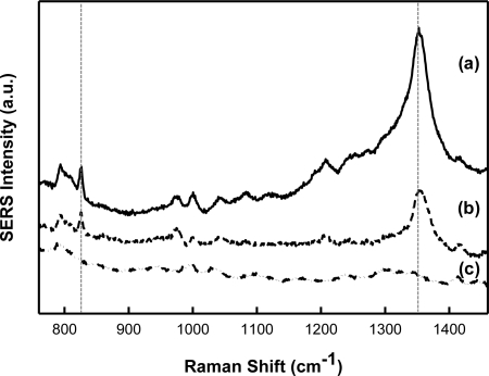
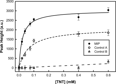
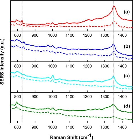
We report on a new sensor strategy that integrates molecularly imprinted polymers (MIPs) with surface enhanced Raman scattering (SERS). The sensor was developed to detect the explosive, 2,4,6-trinitrotoluene (TNT). Micron thick films of sol gel-derived xerogels were deposited on a SERS-active surface as the sensing layer. Xerogels were molecularly imprinted for TNT using non-covalent interactions with the polymer matrix. Binding of the TNT within the polymer matrix results in unique SERS bands, which allow for detection and identification of the molecule in the MIP. This MIP-SERS sensor exhibits an apparent dissociation constant of (2.3 ± 0.3) × 10(-5) M for TNT and a 3 μM detection limit. The response to TNT is reversible and the sensor is stable for at least 6 months. Key challenges, including developing a MIP formulation that is stable and integrated with the SERS substrate, and ensuring the MIP does not mask the spectral features of the target analyte through SERS polymer background, were successfully met. The results also suggest the MIP-SERS protocol can be extended to other target analytes of interest.
Keywords: explosives detection; molecular imprinting; sensor; surface enhanced Raman scattering.
Figures
References
-
- Narayanan A, Varnavski OP, Swager TM, Goodson T. Multiphoton fluorescence quenching of conjugated polymers for TNT detection. J. Phys. Chem. C. 2008;112:881–884.
-
- Yang JS, Swager TM. Fluorescent porous polymer films as TNT chemosensors: Electronic and structural effects. J. Am. Chem. Soc. 1998;120:11864–11873.
-
- Yang JS, Swager TM. Porous shape persistent fluorescent polymer films: An approach to TNT sensory materials. J. Am. Chem. Soc. 1998;120:5321–5322.
-
- Pinnaduwage LA, Yi D, Tian F, Thundat T, Lareau RT. Adsorption of trinitrotoluene on uncoated silicon microcantilever surfaces. Langmuir. 2004;20:2690–2694. - PubMed
-
- Pinnaduwage LA, Wig A, Hedden DL, Gehl A, Yi D, Thundat T, Lareau RT. Detection of trinitrotoluene via deflagration on a microcantilever. J. Appl. Phys. 2004;95:5871–5875.
Publication types
MeSH terms
Substances
LinkOut - more resources
Full Text Sources
Other Literature Sources
Miscellaneous
