. 2011;11(9):8395-411.
doi: 10.3390/s110908395. Epub 2011 Aug 29.

Development of an integrated microfluidic perfusion cell culture system for real-time microscopic observation of biological cells

Affiliations

Development of an integrated microfluidic perfusion cell culture system for real-time microscopic observation of biological cells

Lung Lin Jr et al. Sensors (Basel). 2011.

Abstract

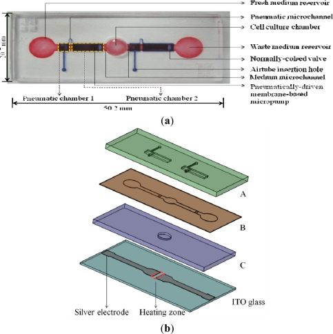
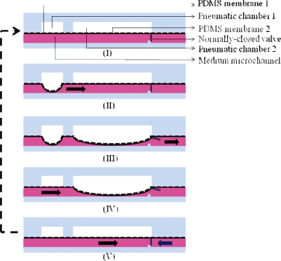
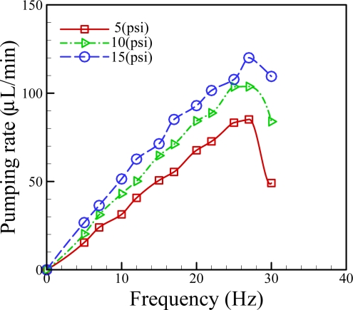
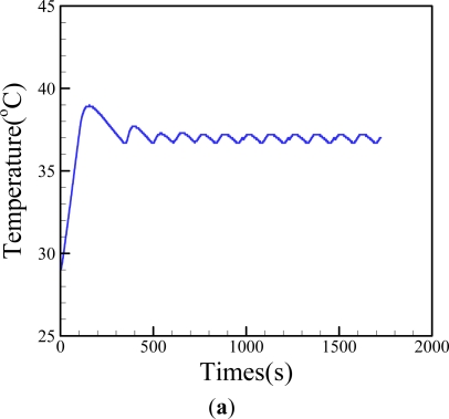
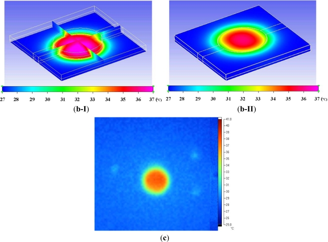
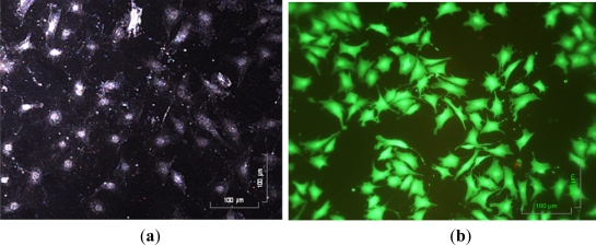
This study reports an integrated microfluidic perfusion cell culture system consisting of a microfluidic cell culture chip, and an indium tin oxide (ITO) glass-based microheater chip for micro-scale perfusion cell culture, and its real-time microscopic observation. The system features in maintaining both uniform, and stable chemical or thermal environments, and providing a backflow-free medium pumping, and a precise thermal control functions. In this work, the performance of the medium pumping scheme, and the ITO glass microheater were experimentally evaluated. Results show that the medium delivery mechanism was able to provide pumping rates ranging from 15.4 to 120.0 μL·min(-1). In addition, numerical simulation and experimental evaluation were conducted to verify that the ITO glass microheater was capable of providing a spatially uniform thermal environment, and precise temperature control with a mild variation of ±0.3 °C. Furthermore, a perfusion cell culture was successfully demonstrated, showing the cultured cells were kept at high cell viability of 95 ± 2%. In the process, the cultured chondrocytes can be clearly visualized microscopically. As a whole, the proposed cell culture system has paved an alternative route to carry out real-time microscopic observation of biological cells in a simple, user-friendly, and low cost manner.

Keywords: ITO glass; cell culture; microfluidics; microheaters; micropumps.


Figures

Figure 1.
Figure 1.
(a) Top view layout (photograph) of the microfluidic cell culture chip; and (b) The assembly of the integrated microfluidic perfusion cell culture system; A–C: microfabricated PDMS plates, and a silver electrode-patterned ITO glass layer.
Figure 2.
Figure 2.
Schematic representation of the medium pumping mechanism using the integrated pneumatically-driven membrane-based micropump coupled with a normally-closed valve (the cross-sectional view) (the arrows indicate the components in the medium pumping mechanism). (I) the rest state, (II) the deformation of PDMS membrane 1 when its corresponding pneumatic chamber 1 above is pressurized, (III) the deformation of PDMS membrane 2 when its corresponding pneumatic chamber 2 is pressurized that also leads to the open of the normally-closed valve, (IV) the deformed PDMS membrane 1 regains to its rest state when the pneumatic chamber 1 is depressurized, and (V) the deformed PDMS membrane 2 regains to its rest state when the pneumatic chamber 2 is depressurized that also leads to the close of the normally-closed valve.
Figure 3.
Figure 3.
(a) Schematic illustration of the fabrication process of ITO-glass microheater chip; (I) the mask with a desirable pattern was attached on a ITO-glass substrate and followed by print screening silver metal paste on it, (II) after removing the mask, the silver electrodes-patterned ITO-glass substrate was created, (III) a 15 μm-thick insulation layer was then screen-printed on the silver electrodes-patterned ITO-glass substrate, and (IV) the fabriacted ITO-glass microheater chip, and (b) A photograph of the ITO-glass microheater chip.
Figure 3.
Figure 3.
(a) Schematic illustration of the fabrication process of ITO-glass microheater chip; (I) the mask with a desirable pattern was attached on a ITO-glass substrate and followed by print screening silver metal paste on it, (II) after removing the mask, the silver electrodes-patterned ITO-glass substrate was created, (III) a 15 μm-thick insulation layer was then screen-printed on the silver electrodes-patterned ITO-glass substrate, and (IV) the fabriacted ITO-glass microheater chip, and (b) A photograph of the ITO-glass microheater chip.
Figure 4.
Figure 4.
Liquid pumping rate profiles of the integrated pneumatic micropump at various applied pneumatic pressures (5, 10, and 15 psi) and frequencies (5–30 Hz).
Figure 5.
Figure 5.
(a) Observation on the temperature profile over time (the set temperature was 37 °C and the temperature variation was evaluated to be within ±0.3 °C); (b) Numerical simulation-based evaluation of the temperature distributions in the (I) cell culture chamber and (II) on the microfluidic cell culture chip; and (c) 2-dimensional thermal IR images on the microfluidic cell chip at the set temperature of 37 °C (top-side view; the circular area represents the cell culture chamber area).
Figure 5.
Figure 5.
(a) Observation on the temperature profile over time (the set temperature was 37 °C and the temperature variation was evaluated to be within ±0.3 °C); (b) Numerical simulation-based evaluation of the temperature distributions in the (I) cell culture chamber and (II) on the microfluidic cell culture chip; and (c) 2-dimensional thermal IR images on the microfluidic cell chip at the set temperature of 37 °C (top-side view; the circular area represents the cell culture chamber area).
Figure 6.
Figure 6.
(a) The observation of articular chondrocyte morphology during cell culture period using a dark field microscope; and (b) the observation of cell viability after 3 day perfusion cell culture using the Live/Dead® fluorescent dye and fluorescent microscope (Green and red dots represent live and dead cells, respectively).

Similar articles

Cited by

References

    1. Xu X, Urban JPG, Browning JA, Tirlapur U, Wilkins RJ, Wu MH, Cui Z, Cui ZF. Influences of buffer systems on chondrocyte growth during long-term culture in alginate. Osteoarthritis Cartilage. 2007;15:396–402. - PubMed
    1. Wu MH, Urban JPG, Cui ZF, Cui Z, Xu X. Effect of extracellular pH on matrix synthesis by chondrocytes in 3D agarose gel. Biotechnol. Prog. 2007;23:430–434. - PubMed
    1. Wu MH, Huang SB, Cui ZF, Cui Z, Lee GB. Development of perfusion-based micro 3-D cell culture platform and its application for high throughput drug testing. Sens. Actuat. B. 2008;129:231–240.
    1. Cui ZF, Xu X, Trainor N, Triffitt JT, Urban JP, Tirlapur UK. Application of multiple parallel perfused microbioreactors and three-dimensional stem cell culture for toxicity testing. Toxicol. In Vitro. 2007;21:1318–1324. - PubMed
    1. Wu MH, Kuo CY. Application of high throughput perfusion micro 3-D cell culture platform for the precise study of cellular responses to extracellular conditions-effect of serum concentrations on the physiology of articular chondrocytes. Biomed. Microdevices. 2011;13:131–141. - PubMed

Publication types

MeSH terms