. 2011 Dec 5:5:131.
doi: 10.3389/fnins.2011.00131. eCollection 2011.

Neurosteroids and GABA(A) Receptor Interactions: A Focus on Stress

Affiliations

Neurosteroids and GABA(A) Receptor Interactions: A Focus on Stress

Benjamin G Gunn et al. Front Neurosci. .

Abstract

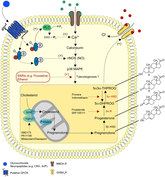
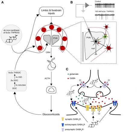
Since the pioneering discovery of the rapid CNS depressant actions of steroids by the "father of stress," Hans Seyle 70 years ago, brain-derived "neurosteroids" have emerged as powerful endogenous modulators of neuronal excitability. The majority of the intervening research has focused on a class of naturally occurring steroids that are metabolites of progesterone and deoxycorticosterone, which act in a non-genomic manner to selectively augment signals mediated by the main inhibitory receptor in the CNS, the GABA(A) receptor. Abnormal levels of such neurosteroids associate with a variety of neurological and psychiatric disorders, suggesting that they serve important physiological and pathophysiological roles. A compelling case can be made to implicate neurosteroids in stress-related disturbances. Here we will critically appraise how brain-derived neurosteroids may impact on the stress response to acute and chronic challenges, both pre- and postnatally through to adulthood. The pathological implications of such actions in the development of psychiatric disturbances will be discussed, with an emphasis on the therapeutic potential of neurosteroids for the treatment of stress-associated disorders.

Keywords: HPA axis; allopregnanolone; anxiety; maternal care; synaptic inhibition.


Figures

Figure 1
Figure 1
A model for the regulation of neurosteroidogenesis. A diagrammatic representation of the pathways for the biosynthesis of 5α3α-THPROG from cholesterol. Receptors and signaling mechanisms potentially regulating steroidogenesis and sites of action of ligands used to target selective enzymatic pathways are additionally illustrated. In the proposed model, NMDA receptor activation may stimulate neurosteroidogenesis on a rapid time scale (Tokuda et al., 2011), possibly via the Ca2+ induced activation of nNOS and p38 MAPK (Izumi et al., 2008). In addition, glucocorticoids may regulate neurosteroidogenesis via rapid non-genomic effects through the activation of Gs-γβ, an effect that stimulates nNOS (Di et al., 2009). Furthermore, a number of stress-related neuropeptides (e.g., CRH, AVP), which are known to signal through Gs-coupled receptor complexes may modulate nNOS activity and thus influence neurosteroid synthesis. Receptors for these neuropeptides can additionally couple to Gq-proteins to raise intracellular Ca2+ levels and thus, couple to signaling cascades similar to those associated with NMDA receptor activation. These GPCR-dependent signaling pathways may provide a functional regulatory link between the stress axis and the de novo synthesis of 5α3α-THPROG.
Figure 2
Figure 2
A model for neurosteroid actions at the GABAA receptor within the stress neurocircuitry. (A) A diagrammatic representation of the HPA axis. The release of CRH from the dorsal–medial parvocellular neurons (mpd) of the PVN is regulated by humoral negative feedback pathways and neuronal inputs from higher brain structures (e.g., the limbic system and the forebrain regions). Inputs from the forebrain and the limbic structures do not directly innervate the PVN, but form polysynaptic connections via a number of predominantly GABAergic “relay nuclei” (red) surrounding the PVN, such as the BST mPOA, DH, and peri-PVN (Cullinan et al., 2008). In the proposed model stress-induced endogenous neurosteroids derived from peripheral sources (e.g., 5α3α-THDOC) and or synthesized de novo within the CNS (e.g., 5α3α-THPROG) may act to enhance the GABAergic inhibition in the PVN and surrounding neurons. Additionally, neurosteroids may modulate inhibitory transmission within limbic and forebrain regions to influence their neuronal output and hence HPA activity. (B) Physiological concentrations of 5α3α-THPROG (100 nM) dramatically reduce the frequency of action currents recorded from WT mpd neurons in a hypothalamic slice preparation containing the PVN and surrounding structures, thus inhibiting their output (i.e., CRH release). Local excitatory inputs (e.g., glutamatergic, green), which are themselves constrained by inhibitory GABAegic neuron (red) originating in the BST and local hypothalamic nuclei (Herman et al., 2002) drive the release of neuropeptides from mpd neuron (gray) of the PVN. (C) A diagrammatic representation of a representative PVN synapse (either GABAergic or glutamatergic). Neurosteroids (e.g., 5α3α-THPROG) synthesized following a stressful challenge, may act both at presynaptic or postsynaptic locations in an autocrine or paracrine fashion respectively to enhance GABAAR inhibition. Note that at presynaptic terminals due to the intraneuronal Cl gradient, GABA may be depolarizing (Trigo et al., 2008) and thus neurosteroids may act to increase neurotransmitter release. Note that although extrasynaptic δ-GABAA receptors are absent in the PVN, other γ2-GABAARs may be expressed extrasynaptically at GABAergic or glutamatergic synapses.

Similar articles

Cited by

References

    1. Agis-Balboa R. C., Pinna G., Zhubi A., Maloku E., Veldic M., Costa E., Guidotti A. (2006). Characterization of brain neurons that express enzymes mediating neurosteroid biosynthesis. Proc. Natl. Acad. Sci. U.S.A. 103, 14602–1460710.1073/pnas.0606544103 - DOI - PMC - PubMed
    1. Akk G., Li P., Bracamontes J., Reichert D. E., Covey D. F., Steinbach J. H. (2008). Mutations of the GABA-A receptor α1 subunitM1 domain reveal unexpected complexity for modulation by neuroactive steroids. Mol. Pharmacol. 74, 614–62710.1124/mol.108.048520 - DOI - PMC - PubMed
    1. Akk G., Shu H. J., Wang C., Steinbach J. H., Zorumski C. F., Covey D. F., Mennerick S. (2005). Neurosteroid access to the GABAA receptor. J. Neurosci. 25, 11605–1161310.1523/JNEUROSCI.4173-05.2005 - DOI - PMC - PubMed
    1. Alfarez D. N., Joels M., Krugers H. J. (2003). Chronic unpredictable stress impairs long-term potentiation in rat hippocampal CA1 area and dentate gyrus in vitro. Eur. J. Neurosci. 17, 1928–193410.1046/j.1460-9568.2003.02622.x - DOI - PubMed
    1. Alfarez D. N., Wiegert O., Joels M., Krugers H. J. (2002). Corticosterone and stress reduce synaptic potentiation in mouse hippocampal slices with mild stimulation. Neuroscience 115, 1119–112610.1016/S0306-4522(02)00483-9 - DOI - PubMed

LinkOut - more resources