

Serum response factor mediated gene activity in physiological and pathological processes of neuronal motility
- PMID: 22164132
- PMCID: PMC3230857
- DOI: 10.3389/fnmol.2011.00049
Serum response factor mediated gene activity in physiological and pathological processes of neuronal motility
Abstract
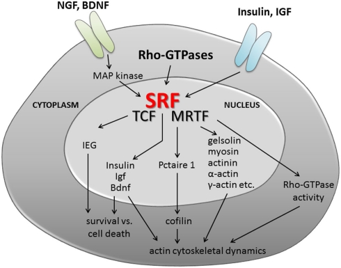
In recent years, the transcription factor serum response factor (SRF) was shown to contribute to various physiological processes linked to neuronal motility. The latter include cell migration, axon guidance, and, e.g., synapse function relying on cytoskeletal dynamics, neurite outgrowth, axonal and dendritic differentiation, growth cone motility, and neurite branching. SRF teams up with myocardin related transcription factors (MRTFs) and ternary complex factors (TCFs) to mediate cellular actin cytoskeletal dynamics and the immediate-early gene (IEG) response, a bona fide indicator of neuronal activation. Herein, I will discuss how SRF and cofactors might modulate physiological processes of neuronal motility. Further, potential mechanisms engaged by neurite growth promoting molecules and axon guidance cues to target SRF's transcriptional machinery in physiological neuronal motility will be presented. Of note, altered cytoskeletal dynamics and rapid initiation of an IEG response are a hallmark of injured neurons in various neurological disorders. Thus, SRF and its MRTF and TCF cofactors might emerge as a novel trio modulating peripheral and central axon regeneration.
Keywords: IEG; MRTF; SRF; TCF; axon; cytoskeleton; neurite; regeneration.
Figures
Similar articles
-
Regulation of Dendritic Synaptic Morphology and Transcription by the SRF Cofactor MKL/MRTF.Front Mol Neurosci. 2021 Nov 2;14:767842. doi: 10.3389/fnmol.2021.767842. eCollection 2021. Front Mol Neurosci. 2021. PMID: 34795561 Free PMC article. Review.
-
A nuclear actin function regulates neuronal motility by serum response factor-dependent gene transcription.J Neurosci. 2009 Apr 8;29(14):4512-8. doi: 10.1523/JNEUROSCI.0333-09.2009. J Neurosci. 2009. PMID: 19357276 Free PMC article.
-
Rho-actin signaling to the MRTF coactivators dominates the immediate transcriptional response to serum in fibroblasts.Genes Dev. 2014 May 1;28(9):943-58. doi: 10.1101/gad.239327.114. Epub 2014 Apr 14. Genes Dev. 2014. PMID: 24732378 Free PMC article.
-
Proteomic analysis of SRF associated transcription complexes identified TFII-I as modulator of SRF function in neurons.Eur J Cell Biol. 2016 Jan;95(1):42-56. doi: 10.1016/j.ejcb.2015.11.002. Epub 2015 Nov 10. Eur J Cell Biol. 2016. PMID: 26589769
-
MKLs: co-factors of serum response factor (SRF) in neuronal responses.Int J Biochem Cell Biol. 2012 Sep;44(9):1444-7. doi: 10.1016/j.biocel.2012.05.008. Epub 2012 May 22. Int J Biochem Cell Biol. 2012. PMID: 22626970 Review.
Cited by
-
Brain-Gut and Microbiota-Gut-Brain Communication in Type-2 Diabetes Linked Alzheimer's Disease.Nutrients. 2024 Aug 3;16(15):2558. doi: 10.3390/nu16152558. Nutrients. 2024. PMID: 39125436 Free PMC article. Review.
-
The SETD8/ELK1/bach1 complex regulates hyperglycaemia-mediated EndMT in diabetic nephropathy.J Transl Med. 2022 Mar 29;20(1):147. doi: 10.1186/s12967-022-03352-4. J Transl Med. 2022. PMID: 35351142 Free PMC article.
-
Intrinsic mechanisms of neuronal axon regeneration.Nat Rev Neurosci. 2018 Jun;19(6):323-337. doi: 10.1038/s41583-018-0001-8. Nat Rev Neurosci. 2018. PMID: 29666508 Free PMC article. Review.
-
Transcriptomic changes during regeneration of the central nervous system in an echinoderm.BMC Genomics. 2014 May 12;15:357. doi: 10.1186/1471-2164-15-357. BMC Genomics. 2014. PMID: 24886271 Free PMC article.
-
Regulation of Dendritic Synaptic Morphology and Transcription by the SRF Cofactor MKL/MRTF.Front Mol Neurosci. 2021 Nov 2;14:767842. doi: 10.3389/fnmol.2021.767842. eCollection 2021. Front Mol Neurosci. 2021. PMID: 34795561 Free PMC article. Review.
References
-
- Alberti S., Krause S. M., Kretz O., Philippar U., Lemberger T., Casanova E., Wiebel F. F., Schwarz H., Frotscher M., Schutz G., Nordheim A. (2005). Neuronal migration in the murine rostral migratory stream requires serum response factor. Proc. Natl. Acad. Sci. U.S.A. 102, 6148–615310.1073/pnas.0501191102 - DOI - PMC - PubMed
LinkOut - more resources
Full Text Sources
Miscellaneous