. 2011;6(12):e28169.
doi: 10.1371/journal.pone.0028169. Epub 2011 Dec 2.

Glycosylation of erythrocyte spectrin and its modification in visceral leishmaniasis

Affiliations

Glycosylation of erythrocyte spectrin and its modification in visceral leishmaniasis

Sajal Samanta et al. PLoS One. 2011.

Abstract

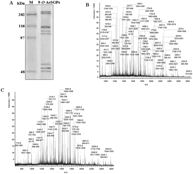
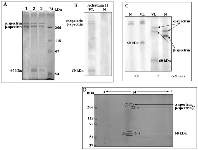
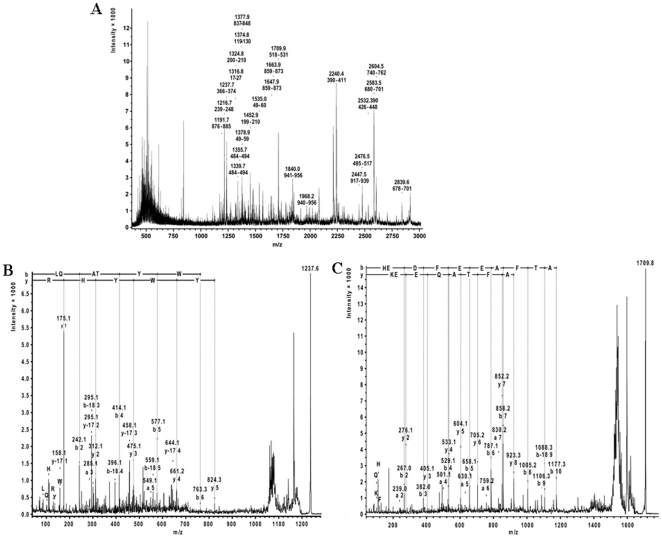
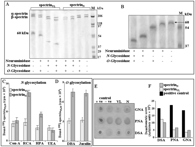
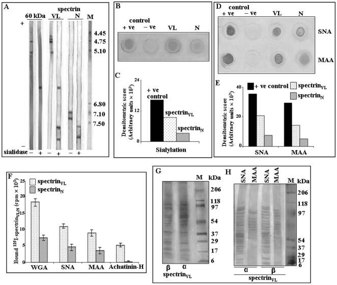
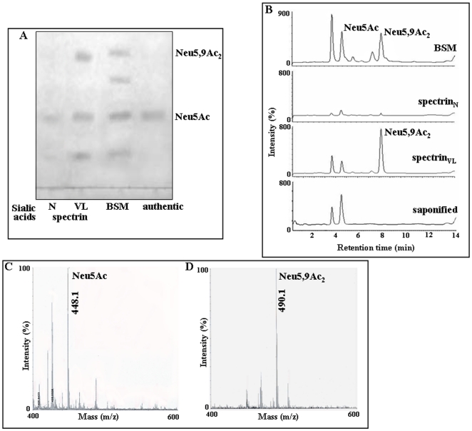
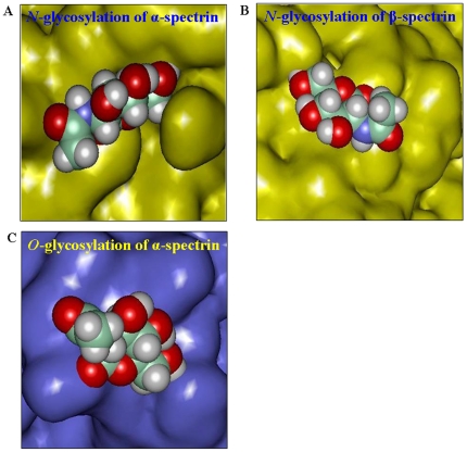
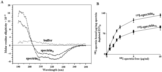
Using a lectin, Achatinin-H, having preferential specificity for glycoproteins with terminal 9-O-acetyl sialic acid derivatives linked in α2-6 linkages to subterminal N-acetylgalactosamine, eight distinct disease-associated 9-O-acetylated sialoglycoproteins was purified from erythrocytes of visceral leishmaniaisis (VL) patients (RBC(VL)). Analyses of tryptic fragments by mass spectrometry led to the identification of two high-molecular weight 9-O-acetylated sialoglycoproteins as human erythrocytic α- and β-spectrin. Total spectrin purified from erythrocytes of VL patients (spectrin(VL)) was reactive with Achatinin-H. Interestingly, along with two high molecular weight bands corresponding to α- and β-spectrin another low molecular weight 60 kDa band was observed. Total spectrin was also purified from normal human erythrocytes (spectrin(N)) and insignificant binding with Achatinin-H was demonstrated. Additionally, this 60 kDa fragment was totally absent in spectrin(N). Although the presence of both N- and O-glycosylations was found both in spectrin(N) and spectrin(VL), enhanced sialylation was predominantly induced in spectrin(VL). Sialic acids accounted for approximately 1.25 kDa mass of the 60 kDa polypeptide. The demonstration of a few identified sialylated tryptic fragments of α- and β-spectrin(VL) confirmed the presence of terminal sialic acids. Molecular modelling studies of spectrin suggest that a sugar moiety can fit into the potential glycosylation sites. Interestingly, highly sialylated spectrin(VL) showed decreased binding with spectrin-depleted inside-out membrane vesicles of normal erythrocytes compared to spectrin(N) suggesting functional abnormality. Taken together this is the first report of glycosylated eythrocytic spectrin in normal erythrocytes and its enhanced sialylation in RBC(VL). The enhanced sialylation of this cytoskeleton protein is possibly related to the fragmentation of spectrin(VL) as evidenced by the presence of an additional 60 kDa fragment, absent in spectrin(N) which possibly affects the biology of RBC(VL) linked to both severe distortion of erythrocyte development and impairment of erythrocyte membrane integrity and may provide an explanation for their sensitivity to hemolysis and anemia in VL patients.


Conflict of interest statement

Competing Interests: The authors have declared that no competing interests exist.

Figures

Figure 1
Figure 1. Purification of 9-O-AcSGPs and identification of spectrin.
A. A representative SDS-PAGE (7.5%) profile of purified 9-O-AcSGPs from RBCVL. Lane M shows molecular weight standards. B–C. PMF spectra of tryptic fragments of two high molecular weight 9-O-AcSGPs were identified as α- (B) and β-spectrin (C) by MALDI-TOF MS. Each fragment is denoted by their m/z values and sequence range in human α- and β-spectrin.
Figure 2
Figure 2. Purification and characterization of spectrin.
A. Purification of spectrinVL and spectrinN. A representative SDS-PAGE (7.5%) profile of SpectrinN (2.0 µg, lane 1) and spectrinVL (2.0 µg, lane 2), purified from RBCN and RBCVL as described by Ungewickell et al . Purified spectrinVL was further passed through an Achatinin-H-Sepharose 4B affinity column and 9-O-acetylated sialic acid containing spectrinVL (2.0 µg, lane 3) was purified as described in Materials and Methods. Lane M shows molecular weight standards. B. Presence of 9-O-AcSA as detected by Western blot analysis. Equal amounts (2 µg) of purified spectrinVL and spectrinN were transferred onto nitrocellulose membrane after SDS-PAGE (8.5%). The blots were incubated overnight at 4°C with Achatinin-H and processed as described in Materials and Methods. C. Equal amount (2 µg) of purified spectrinVL and spectrinN were separated both on 5 and 7.5% SDS-PAGE under similar conditions. D. Two dimensional (2D) gel electrophoresis of spectrinVL. A representative 2D (pI range 4–7, 4–15% gradient) profile of purified spectrin (100 µg) from RBCVL after staining with Coomassie is shown.
Figure 3
Figure 3. Identification of 60 kDa band.
A. The PMF spectra of tryptic fragments of 60 kDa glycoprotein. PMF spectra of tryptic fragments of 60 kDa were identified as N-terminal fragment of α-spectrin by MALDI-TOF MS. Each fragment is denoted by their m/z values and sequence range within the 955 amino acids of human α-spectrin (marked with yellow in Fig. S1). B–C. Confirmation of the sequence of the identified tryptic fragments by MALDI-TOF-TOF mass spectrometry. The MS/MS spectrum was analyzed with database-dependent MASCOT as well as database-independent Sequit! software systems yielding the same results. Two representative PSD spectra of the MS/MS analysis of the fragment (B) LQATYWYHR (m/z = 1237.6) and (C) HEDFEEAFTAQEEK (m/z = 1237.6) of α-spectrin and SGP-60. The N and C terminal fragment ions are denoted according to standard nomenclature and immonium ions displayed in single amino acid code.
Figure 4
Figure 4. Demonstration of N-and O-glycosylation.
A. Demonstration of N-and O-glycosylation of spectrin by enzyme deglycosylation. Equal amount (5 µg) of purified spectrinVL and spectrinN was treated with neuraminidase from Arthrobacter ureafaciens to remove the terminal sialic acids and subsequently desialylated spectrinVL and spectrinN was incubated separately with N-glycosidase F, O-glycosidase or a combination of N- and O-glycosidase as indicated. SpectrinVL/N before and after the respective enzyme treatments were analyzed by SDS-PAGE as described in Materials and Methods. B. Demonstration of sialylation, N- and O-glycosylation in 60 kDa fragment. Gel-eluted purified 60 kDa fragment (1.0 µg) was initially desialylated with Arthrobacter ureafaciens neuraminidase overnight at 37°C. Subsequently the desialylated 60 kDa fragment was treated separately with N-glycosidase F, O-glycosidase F or a combination of both and analyzed by SDS-PAGE (7.5%) along with the untreated protein as described in Materials and Methods. Gel was stained with silver staining method. Lane M shows molecular weight standards. C–D. Demonstration of N- and O-glycosylation by lectin binding with 125I-spectrinVL/N. Fixed concentrations of 125I-spectrinVL/N were processed separately to demonstrate their binding with several Sepharose/agarose bound ConA, RCA, HPA, UEA, DBA and Jacalin lectins (25 µl bead volume) having different sugar-linkage specificity as described in Materials and Methods. E–F. Demonstration of N- and O-glycosylation by lectin binding with DIG-glycan. E. Equal amount (2.0 µg) of spectrinVL and spectrinN was dot blotted on NC-paper and analyzed by DIG-glycan and differentiation kit using several lectins (GNA, PNA, DSA) following manufacturer's protocol. F. Representative bar graph of densitometric scores of corresponding spots.
Figure 5
Figure 5. Presence of Neu5Ac and Neu5,9Ac2 in spectrinVL by biochemical methods.
A. Enhanced sialylation demonstrated by IEF. Equal amounts (3.0 µg) of purified spectrinVL, 60 kDa band and spectrinN before and after removal of sialic acids were analyzed by IEF within a pH gradient of 3–10 and the respective bands visualized by silver staining. Lane M shows the pI markers. B–C. Enhanced sialylation in spectrinVL. Equal amount (1.0 µg) of purified spectrinVL and spectrinN was analyzed by using DIG-glycan detection kits and total sialylation was compared based on the densitometric scores of spots (B). Representative bar graph of densitometric scores of corresponding spots (C). D–E. Detection of linkage-specific terminal sialic acids in spectrinVL. Equal amount (2.0 µg) of spectrinVL and spectrinN was dot blotted on NC-paper and analyzed by DIG glycan and differentiation kit using SNA and MAA lectins following manufacturer's protocol (D). Densitometric scores of corresponding spots are shown as bar graph (E). F. Binding of 125I-spectrinVL/N with several sialic acid binding lectins. To demonstrate the presence or absence of terminal sialic acids, a fixed concentrations of 125I-spectrinVL/N were analyzed by binding with Sepharose/agarose bound WGA, SNA, MAA, Achatinin-H (25 µl bead volume) having specificity towards linkage specific sialic acids as described in materials and methods. Bound radioactivity of 125I-spectrinVL/N was measured by Gamma-counter and represented as bar graphs. G–H. Detection of sialylated tryptic fragments in spectrinVL. The α and β subunits of purified spectrinVL were digested separately by restricted amount of trypsin. Such controlled digested and extracted tryptic fragments were dried and redissolved and an aliquot was separated in SDS-PAGE (7.5%–15% gradient) (G). Subsequently the presence of sialic acids on resulting tryptic fragments was analyzed by binding with SNA-agarose and MAA-agarose separately and followed by electrophoresis on SDS-PAGE (7.5%–15% gradient) (H) as described in Materials and Methods. Lane M shows the molecular weight standerds.
Figure 6
Figure 6. Presence of Neu5Ac and Neu5,9Ac2 in spectrinVL by analytical methods.
A. Thin layer chromatography (TLC). Glycosidically bound sialic acids of spectrinVL were subjected to acid hydrolysis, purified, separated on a TLC plate and detected by staining with orcinol/HCl spray reagent and baking at 180°C. Similarly processed free sialic acids released from BSM served as standard. Additionally, commercially available Neu5Ac was used as references. For comparison liberated sialic acids from purified spectrinN were similarly analyzed. B. Enhanced presence of Neu5Ac and Neu5,9Ac2 in spectrinVL as determined by fluorimetric HPLC. Glycosidically bound sialic acids released from spectrinVL by acid hydrolysis were derivatized with DMB and analyzed by fluorimetric HPLC before and after saponification as described in Materials and Methods. A representative chromatogram of the spectrinVL and spectrinN derived sialic acids showed the presence of fluorescent derivatives of free sialic acids. In parallel sialic acids of BSM similarly analyzed under identical conditions served as standard. C–D. Identification of sialic acids by MALDI-TOF MS. Fractions corresponding to peaks of Neu5Ac (C) and Neu5,9Ac2 (D) were collected after fluorimetric HPLC, spotted and analyzed by MALDI-TOF MS using DHBA matrix as described in Materials and Methods. Positive ion mode was used for mass-spectrometric analysis with 1000 laser shots per spot.
Figure 7
Figure 7. Space filling structural representation of GlcNAc in spectrinN.
Sugar moiety are colored by atoms (C = green, O = red, N = blue and H = white). The protein model is represented as conolly surface. A. N- glycosylation of α-spectrin is shown in yellow color at position Asn-1625. B. N- glycosylation of β-spectrin is shown in yellow color at position Asn-194. C. O- glycosylation of α-spectrin is shown in blue color at position Thr-817.
Figure 8
Figure 8. Physicochemical study of structural modification of spectrinVL.
A. CD-spectra. Far-UV CD spectra of spectrinVL and spectrinN in phosphate buffer (20 mM, pH 7.0) indicating the molar residue ellipticity as a function of wavelength along with the buffer only. B. Binding of 125I-spectrin to spectrin-depleted IOVN. Various concentrations of 125I-spectrinVL/N were incubated with a constant amount of spectrin-depleted-IOVN followed by determination of specific binding as described in Materials and Methods.

Similar articles

Cited by

References

    1. Bennett V, Baines AJ. Spectrin and ankyrin-based pathways: metazoan inventions for integrating cells into tissues. Physiol Rev. 2001;81:1353–1392. - PubMed
    1. Bossi D, Russo M. Hemolytic anemias due to disorders of red cell membrane skeleton. Mol Aspects Med. 1996;17:171–188. - PubMed
    1. Resmi H, Pekçetin Ç, Güner G. Erythrocyte membrane and cytoskeletal protein glycation and oxidation in short-term diabetic rabbits. Clin Exp Med. 2001;1:187–193. - PubMed
    1. Starodubtseva MN, Kuznetsova TG, Yegorenkov NI, Cherenkevich SN. Structural and mechanical characteristics of erythrocyte membranes in patients with type 2 diabetes mellitus. Bull Exp Biol Med. 2008;145:99–103. - PubMed
    1. Schauer R. Sialic acids as regulators of molecular and cellular interactions. Curr Opin Struct Biol. 2009;19:507–514. - PMC - PubMed

Publication types

MeSH terms