. 2011;6(12):e28295.
doi: 10.1371/journal.pone.0028295. Epub 2011 Dec 2.

Monkeypox disease transmission in an experimental setting: prairie dog animal model

Affiliations

Monkeypox disease transmission in an experimental setting: prairie dog animal model

Christina L Hutson et al. PLoS One. 2011.

Abstract

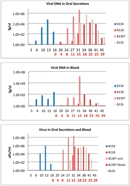
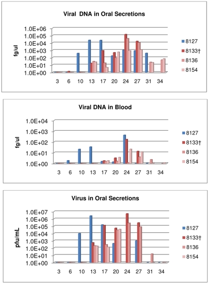
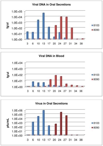
Monkeypox virus (MPXV) is considered the most significant human public health threat in the genus Orthopoxvirus since the eradication of variola virus (the causative agent of smallpox). MPXV is a zoonotic agent endemic to forested areas of Central and Western Africa. In 2003, MPXV caused an outbreak in the United States due to the importation of infected African rodents, and subsequent sequential infection of North American prairie dogs (Cynomys ludovicianus) and humans. In previous studies, the prairie dog MPXV model has successfully shown to be very useful for understanding MPXV since the model emulates key characteristics of human monkeypox disease. In humans, percutaneous exposure to animals has been documented but the primary method of human-to-human MPXV transmission is postulated to be by respiratory route. Only a few animal model studies of MPXV transmission have been reported. Herein, we show that MPXV infected prairie dogs are able to transmit the virus to naive animals through multiple transmission routes. All secondarily exposed animals were infected with MPXV during the course of the study. Notably, animals secondarily exposed appeared to manifest more severe disease; however, the disease course was very similar to those of experimentally challenged animals including inappetence leading to weight loss, development of lesions, production of orthopoxvirus antibodies and shedding of similar levels or in some instances higher levels of MPXV from the oral cavity. Disease was transmitted via exposure to contaminated bedding, co-housing, or respiratory secretions/nasal mucous (we could not definitively say that transmission occurred via respiratory route exclusively). Future use of the model will allow us to evaluate infection control measures, vaccines and antiviral strategies to decrease disease transmission.


Conflict of interest statement

Competing Interests: The authors have declared that no competing interests exist.

Figures

Figure 1
Figure 1. Bedding/fomite group: detection of viral DNA and viable virus from oropharyngeal swabs and blood.
Four primary challenged animals were inoculated via intranasal route with 9×10³ pfu (0.07XLD50) of West African MPXV and placed into one of three experimental groups. For the bedding group, the primary challenged animal (blue bars) was housed for 16 days in a large trough, at which time the challenged animal was removed and three naive animals (red/mauve bars) were placed into the trough for housing. Blood and oral swabs were taken from each animal throughout the study and subsequently tested for viral DNA and live virus. Days are plotted on the X axis in black for day p.i. of primary challenged animal; in red for day post placement in trough. Samples yielding positive DNA (fg/ul) or viable virus (pfu/mL) are plotted on the Y axis in a log scale.
Figure 2
Figure 2. Co-house group: detection of viral DNA and viable virus from oropharyngeal swabs and blood.
Four primary challenged animals were inoculated via intranasal route with 9×10³ pfu (0.07XLD50) of West African MPXV and placed into one of three experimental groups. For the co-housed group; primary challenged animal was challenged with virus and housed in a large trough with 3 naive animals. Blood and oral swabs were taken from each animal throughout the study and subsequently tested for viral DNA and live virus. Days are plotted on the X axis. Samples yielding positive DNA (fg/ul) or viable virus (pfu/mL) are plotted on the Y axis in a log scale.
Figure 3
Figure 3. Respiratory secretions group-1: detection of viral DNA and viable virus from oropharyngeal swabs and blood.
Four primary challenged animals were inoculated via intranasal route with 9×10³ pfu (0.07XLD50) of West African MPXV and placed into one of three experimental groups. For the respiratory secretion group; large metal rabbit cages with holes on one side were utilized to single house animals. Cages were placed approximately one inch apart with holes from the challenged animals' cages facing the holed-side from the naive animals' cages. Blood and oral swabs were taken from each animal throughout the study and subsequently tested for viral DNA and live virus. Days are plotted on the X axis. Samples yielding positive DNA (fg/ul) or viable virus (pfu/mL) are plotted on the Y axis in a log scale.
Figure 4
Figure 4. Respiratory secretions group 1 disease time-line.
Primary challenged animal 8103 was inoculated via intranasal route with 9×10³ pfu (0.07XLD50) of West African MPXV and placed in a large metal cage with holes on one side which faced the holed-side of a cage housing naive animal 8090. Blood , oral swabs, weights and lesion counts were taken from each animal throughout the study. Total time that viable virus and viral DNA were present are illustrated with bars. The initial detection of antibodies, weight loss occurrence and lesion formation are illustrated with arrows. AB: development of antibodies. WL: weight loss >5%.
Figure 5
Figure 5. Respiratory secretions group-2: detection of viral DNA and viable virus from oropharyngeal swabs and blood.
Four primary challenged animals were inoculated via intranasal route with 9×10³ pfu (0.07XLD50) of West African MPXV and placed into one of three experimental groups. For the respiratory secretion group; large metal rabbit cages with holes on one side were utilized to single house animals . Cages were placed approximately one inch apart with holes from the challenged animals' cages facing the holed-side from the naive animals' cages. Blood and oral swabs were taken from each animal throughout the study and subsequently tested for viral DNA and live virus. Days are plotted on the X axis. Samples yielding positive DNA (fg/ul) or viable virus (pfu/mL) are plotted on the Y axis in a log scale.
Figure 6
Figure 6. Average blood chemistry values for prairie dogs.
Four primary challenged animals were inoculated via intranasal route with 9×10³ pfu (0.07XLD50) of West African MPXV and placed into one of three experimental groups with naive animals. Blood was taken from each animal before the study began and at time of euthanasia or study completion. Values are for all animals pre-bleeds (n = 14), post-bleeds for animals that survived infection (n = 9) and animals that were euthanized due to extreme morbidity (n = 3); one of which was prairie dog 8130 (A). Error bars show the standard deviation. Values are grouped according to unit of measurement: U/L (A), MG/DL (B), MMOL/L (C) and G/DL (D).

Similar articles

Cited by

References

    1. Breman JG, Nakano JH, Coffi E, Godfrey H, Gautun JC. Human poxvirus disease after smallpox eradication. Am J Trop Med Hyg. 1977;26:273–281. - PubMed
    1. Hutin YJ, Williams RJ, Malfait P, Pebody R, Loparev VN, et al. Outbreak of human monkeypox, Democratic Republic of Congo, 1996 to 1997. Emerg Infect Dis. 2001;7:434–438. - PMC - PubMed
    1. Khodakevich L, Szczeniowski M, Manbu mD, Jezek Z, Marennikova S, et al. The role of squirrels in sustaining monkeypox virus transmission. Trop Geogr Med. 1987;39:115–122. - PubMed
    1. Centers for Disease Control and Prevention. Update: multistate outbreak of monkeypox–Illinois, Indiana, Kansas, Missouri, Ohio, and Wisconsin, 2003. MMWR Morb Mortal Wkly Rep. 2003;52:561–564. - PubMed
    1. Hutson CL, Lee KN, Abel J, Carroll DS, Montgomery JM, et al. Monkeypox zoonotic associations: insights from laboratory evaluation of animals associated with the multi-state US outbreak. Am J Trop Med Hyg. 2007;76:757–768. - PubMed