. 2011;6(12):e28352.
doi: 10.1371/journal.pone.0028352. Epub 2011 Dec 2.

In vitro model of vascularized bone: synergizing vascular development and osteogenesis

Affiliations

In vitro model of vascularized bone: synergizing vascular development and osteogenesis

Cristina Correia et al. PLoS One. 2011.

Abstract

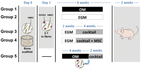
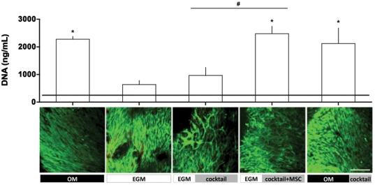
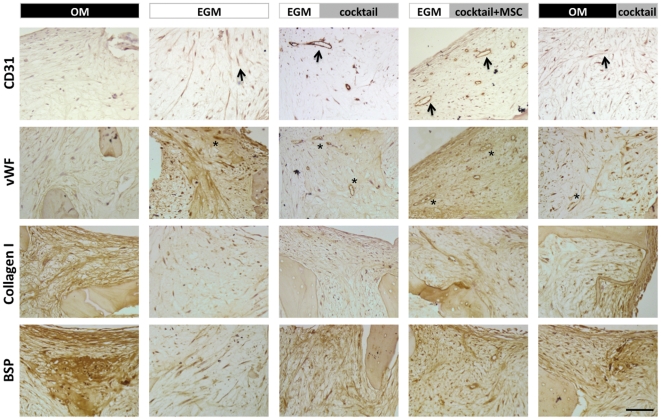
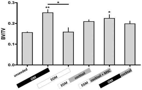
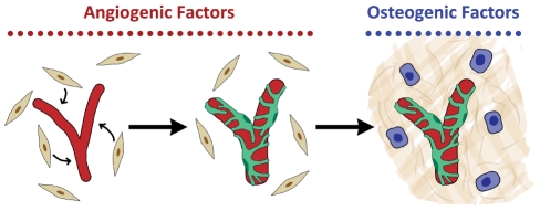
Tissue engineering provides unique opportunities for regenerating diseased or damaged tissues using cells obtained from tissue biopsies. Tissue engineered grafts can also be used as high fidelity models to probe cellular and molecular interactions underlying developmental processes. In this study, we co-cultured human umbilical vein endothelial cells (HUVECs) and human mesenchymal stem cells (MSCs) under various environmental conditions to elicit synergistic interactions leading to the colocalized development of capillary-like and bone-like tissues. Cells were encapsulated at the 1:1 ratio in fibrin gel to screen compositions of endothelial growth medium (EGM) and osteogenic medium (OM). It was determined that, to form both tissues, co-cultures should first be supplied with EGM followed by a 1:1 cocktail of the two media types containing bone morphogenetic protein-2. Subsequent studies of HUVECs and MSCs cultured in decellularized, trabecular bone scaffolds for 6 weeks assessed the effects on tissue construct of both temporal variations in growth-factor availability and addition of fresh cells. The resulting grafts were implanted subcutaneously into nude mice to determine the phenotype stability and functionality of engineered vessels. Two important findings resulted from these studies: (i) vascular development needs to be induced prior to osteogenesis, and (ii) the addition of additional hMSCs at the osteogenic induction stage improves both tissue outcomes, as shown by increased bone volume fraction, osteoid deposition, close proximity of bone proteins to vascular networks, and anastomosis of vascular networks with the host vasculature. Interestingly, these observations compare well with what has been described for native development. We propose that our cultivation system can mimic various aspects of endothelial cell-osteogenic precursor interactions in vivo, and could find utility as a model for studies of heterotypic cellular interactions that couple blood vessel formation with osteogenesis.


Conflict of interest statement

Competing Interests: The authors have declared that no competing interests exist.

Figures

Figure 1
Figure 1. Schematic of experimental approaches.
Groups 1 & 2 are ‘controls’ where constructs were provided osteogenic supplements (OM) or endothelial factors (EGM) for 6 weeks. In Groups 3 & 4, vascular differentiation was induced for 2 weeks before adding osteogenic factors in a cocktail medium (EGM+OM at 1∶1 ratio). No additional cells were added at this point in Group 3 (EGM|cocktail), while osteo-induced MSCs were seeded into the pore spaces in Group 4 (EGM|cocktail+MSCs). These were compared with cultures in Group 5 where only MSCs were added initially and cultured in OM for 4 weeks. A co-culture of HUVECs and MSCs were then added and constructs cultured in cocktail medium (OM|cocktail) for remaining 2 weeks. Constructs of all groups were implaneted sub-cutaneously in nude mice for additional 2 weeks.
Figure 2
Figure 2. DNA contents of constructs after 6 weeks of in vitro culture.
Upper: Horizontal line indicates day 1 values. n = 3; * indicate p<0.05 in comparison to day 1 values. # indicate p<0.05 among groups. Lower: Live/dead imaging of constructs after in vivo culture. Scale bar = 200 µm.
Figure 3
Figure 3. Immunohistological analysis of constructs cultured in vitro.
Engineered bone grafts were evaluated for the expression of vascular and osteogenic proteins. CD31 (black arrows) and vWF (indicated by *) expression was observed in constructs from all groups. Vascular structures were most developed in EGM|cocktail and EGM|cocktail+MSC groups. Collagen I and BSP were readily apparent in the OM group. Expression was observed in individual cells in EGM group, but not distributed through matrix. Both collagen I and BSP were observed in all other groups. In EGM|cocktail+MSC group, BSP appeared in close proximity to the vascular structures. Scale bar = 20 µm.
Figure 4
Figure 4. Ratio of bone material volume over tissue volume (BV/TV) of constructs after in vitro culture.
n = 3; * indicate p<0.05 and ** indicate p<0.001 in comparison to unseeded group. # indicate p<0.05 among both groups.
Figure 5
Figure 5. Composition of engineered grafts.
Top row: Gross images of constructs post-harvest showing the translucent capsule and the in-growth of blood vessels. Constructs are perfused to different extents in different experimental groups. The large blood vessels also seem to anastomose to engineered microvasculature resulting in blood flow through capillary-like networks. This is particularly evident in EGM|cocktail and EGM|cocktail+MSC groups. Scale bar = 500 µm. Second row: Constructs were stained with anti-human CD31. Capsular region filled with mouse cells are shown with asterisks (*). In EGM|cocktail and EGM|cocktail+MSC groups, lumen (stained with anti-human CD31) are larger and well-developed (black arrows) with red blood cells inside (stained with hematoxylin). In OM|cocktail group, there is little evidence of human-derived vascular tissue. Large vessel-like structures in the capsular region are not stained with anti-human CD31 mAb and are probably of mouse origin (yellow arrow heads). Scale bar = 50 µm. Rows 3 and 4: In vivo bone development. Goldner's Masson trichrome staining of non-decalcified constructs indicate distinct levels of osteoid formation among groups (red stains indicated by black arrow heads). Third row: scale bar = 20 µm, Bottom row: scale bar = 500 µm.
Figure 6
Figure 6. Model of in vitro coordination of vascular and bone tissue development.
In a co-culture of ECs and hMSCs (beige, spindle-shaped cells), angiogenic supplements provide cues to stimulate the formation of primitive vascular networks (red) by the ECs (dispersed ECs are not shown in the model). These, in turn, recruit hMSCs into a pericyte-like role (green) that enables the vascular network to remain stable when osteogenic cues are provided. These cues induce osteoblast formation (blue cells) and deposition of mineralized matrix (beige).

Similar articles

Cited by

References

    1. Mikos AG, Herring SW, Ochareon P, Elisseeff J, Lu HH, et al. Engineering complex tissues. Tissue Engineering. 2006;12:3307–3339. - PMC - PubMed
    1. Wang Y, Wan C, Deng LF, Liu XM, Cao XM, et al. The hypoxia-inducible factor a pathway couples angiogenesis to osteogenesis during skeletal development. Journal of Clinical Investigation. 2007;117:1616–1626. - PMC - PubMed
    1. Maes C, Kobayashi T, Selig MK, Torrekens S, Roth SI, et al. Osteoblast Precursors, but Not Mature Osteoblasts, Move into Developing and Fractured Bones along with Invading Blood Vessels. Developmental Cell. 2010;19:329–344. - PMC - PubMed
    1. Hofmann A, Ritz U, Verrier S, Eglin D, Alini M, et al. The effect of human osteoblasts on proliferation and neo-vessel formation of human umbilical vein endothelial cells in a long-term 3D co-culture on polyurethane scaffolds. Biomaterials. 2008;29:4217–4226. - PubMed
    1. Choong CSN, Hutmacher DW, Triffitt JT. Co-culture of bone marrow fibroblasts and endothelial cells on modified polycaprolactone substrates for enhanced potentials in bone tissue engineering. Tissue Engineering. 2006;12:2521–2531. - PubMed

Publication types

Substances