. 2012 Feb 15;442(1):77-84.
doi: 10.1042/BJ20111903.

Staphylococcus aureus DinG, a helicase that has evolved into a nuclease

Affiliations

Staphylococcus aureus DinG, a helicase that has evolved into a nuclease

Anne-Marie McRobbie et al. Biochem J. .

Abstract

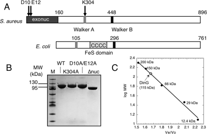
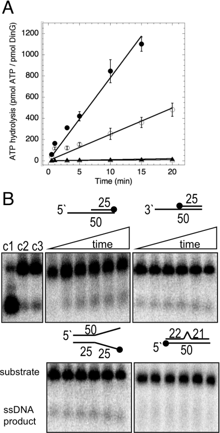
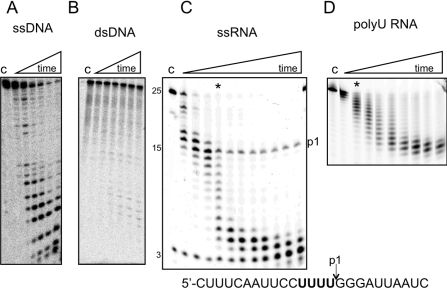
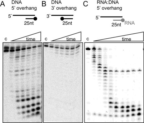
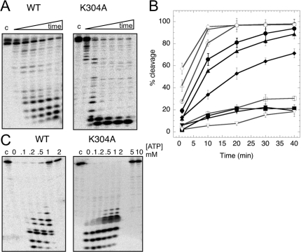
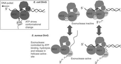
DinG (damage inducible gene G) is a bacterial superfamily 2 helicase with 5′→3′ polarity. DinG is related to the XPD (xeroderma pigmentosum complementation group D) helicase family, and they have in common an FeS (iron–sulfur)-binding domain that is essential for the helicase activity. In the bacilli and clostridia, the DinG helicase has become fused with an N-terminal domain that is predicted to be an exonuclease. In the present paper we show that the DinG protein from Staphylococcus aureus lacks an FeS domain and is not a DNA helicase, although it retains DNA-dependent ATP hydrolysis activity. Instead, the enzyme is an active 3′→5′ exonuclease acting on single-stranded DNA and RNA substrates. The nuclease activity can be modulated by mutation of the ATP-binding cleft of the helicase domain, and is inhibited by ATP or ADP, suggesting a modified role for the inactive helicase domain in the control of the nuclease activity. By degrading rather than displacing RNA or DNA strands, the S. aureus DinG nuclease may accomplish the same function as the canonical DinG helicase.


Figures

Figure 1
Figure 1. Comparison and purification of sarDinG proteins
(A) Comparison of the domain organization of DinG from S. aureus and E. coli, showing the exonuclease (exonuc) domain in the former and the FeS-binding domain in the latter. The positions of the Walker A and Walker B motifs in both proteins are indicated by grey and black boxes respectively. The positions of the three point mutations made in sarDinG are indicated by arrows. (B) SDS/PAGE showing purified WT and mutant sarDinG proteins. The molecular mass in kDa is indicated on the left-hand side. (C) Elution profile of WT sarDinG on a calibrated Superdex 200 gel-filtration column. The standard curve shown was calculated using the elution volumes of a set of standard proteins of known molecular mass, allowing the estimation of the native molecular mass of DinG, which was consistent with a monomeric composition.
Figure 2
Figure 2. ATPase and helicase activity of sarDinG
(A) The rate of ATP hydrolysis by WT and K304A mutant DinG in the presence and absence of ssDNA. WT DinG displayed a modest ATPase activity (kcat=25 min−1) that was stimulated by ssDNA (kcat=78 min−1). The ATPase activity of the K304A mutant was negligible, as expected. Data points represent the mean of triplicate measurements, with S.E.M.s indicated. ● and ○, WT DinG; ▲ and Δ, K304A mutant. Closed symbols indicate the presence of ssDNA, open symboles indicate the the absence of ssDNA. (B) Helicase assays to determine the ability of 1 μM sarDinG to unwind DNA molecules consisting of a 5′-overhang, 3′-overhang, splayed duplex and duplex with an internal 7 nt bubble. The black dots indicate the site of the 5′-32P radioactive label. Protein (1 μM) was incubated with 10 nM radiolabelled DNA at 37°C in helicase buffer and reactions were stopped after 1, 10, 20, 30, 40 and 60 min and analysed by acrylamide gel electrophoresis. No appreciable helicase activity was detected for any substrate. Controls: c1, boiled substrate; c2, no protein control at 60 min; c3, absence of ATP/Mg at 60 min.
Figure 3
Figure 3. Nuclease activity of sarDinG on ssDNA and ssRNA substrates
(A) sarDinG (500 nM) was incubated with 10 nM 32P-labelled oligonucleotide (DNA25X) and 5 mM MgCl2 at 37°C. Progressive cleavage in a 3′→5′ direction was observed. Reactions were stopped after 1, 10, 20, 30, 40 and 60 min and analysed by denaturing acrylamide gel electrophoresis. c, control reaction incubated at 37°C for 60 min in the absence of protein. (B) The activity of sarDinG on a DNA duplex with a central 7 nt bubble. Reaction conditions were identical with those described for (A). No cleavage activity was observed. (C) Cleavage of a 25 nt ssRNA oligonucleotide (RNA25) with a 5′ fluorescein label by sarDinG. Progressive cleavage in a 3′→5′ direction was observed as for ssDNA. A pronounced pause site (p1), which persisted to the end of the reaction time, was observed to correspond to a run of four uracil residues in the RNA sequence (shown in bold below). The reaction conditions were the same as for (A) with time points of 0.5, 1, 2, 5, 10, 15, 20, 30, 60, 90 and 120 min. The 5 min time point is indicated with an asterisk. (D) Cleavage of a 15 nt poly-uracil RNA oligonucleotide (polyU) was carried out as described for (A). Cleavage kinetics were more uniform and considerably lower than those observed in (C), confirming the sequence-dependence of the nuclease reaction. The asterisk indicates the 5 min time point. Time points correspond to incubation for 1, 5, 10, 15, 20, 30, 40, 60 and 90 min.
Figure 4
Figure 4. DinG helicase substrates are degraded by sarDinG
(A) sarDinG (500 nM) was incubated with a DNA duplex with a 5′ overhang, consisting of an unlabelled 50 nt oligonucleotide (DNA50) annealed to a 5′-end labelled oligonucleotide (DNA25H) and 5 mM MgCl2 at 37°C. Progressive cleavage in a 3′→5′ direction was observed. Reactions were stopped after 1, 10, 20, 30, 40 and 60 min and analysed by denaturing acrylamide gel electrophoresis. c, control reaction incubated at 37°C for 60 min in the absence of protein. (B) Reaction as in (A) but with a DNA duplex with a 3′ overhang, consisting of an unlabelled 50 nt oligonucleotide (DNA50) annealed to a 5′-end labelled oligonucleotide (DNA25X). No appreciable cleavage was observed. (C) sarDinG (500 nM) was incubated with an RNA–DNA hybrid molecule consisting of an unlabelled 40 nt oligonucleotide (DNA40) annealed to a 25 nt RNA oligonucleotide with a 5′-fluorecein label (RNA25). Reaction conditions were as for (A) with time points 0.5, 1, 2, 5, 10, 15, 20, 30 and 60 min. The same pause site observed in Figure 3(C) was observed.
Figure 5
Figure 5. Modulation of nuclease activity by the helicase domain and nucleotides
(A) Comparison of the cleavage activity of WT and K304A sarDinG with a 32P-radiolabelled 5′ overhang substrate and 5 mM MgCl2 at 37°C. The K304A mutant displayed significantly higher nuclease activity than the WT enzyme. Reactions were stopped after 1, 10, 20, 30, 40 and 60 min and analysed by denaturing acrylamide gel electrophoresis. c, control reaction incubated at 37°C for 60 min in the absence of protein. (B) Comparison of the activities of the WT and K304A mutant proteins on a range of DNA substrates as a function of time. Substrates tested were ssDNA (● and ○), 5′ overhang (▲ and Δ), 3′ overhang (■ and □), splayed duplex (◆ and ◇) and bubble (▼ and ∇). WT data points have closed symbols, and K304A data points have open symbols. Each data point represents the mean of triplicate experiments with S.E.M.s indicated. (C) Comparison of the effect of ATP on the nuclease activity of WT and K304A sarDinG. Reactions were carried out under standard conditions with 5 mM MgCl2 and a varying concentration of ATP for 60 min using the DNA25X oligonucleotide, and analysed as described previously. WT DinG was inhibited fully by 1 mM ATP, but the K304A mutant was still active in the presence of 2 mM ATP and only inhibited fully by 5 mM ATP.
Figure 6
Figure 6. Cartoon representation of a possible reaction mechanism for sarDinG
E. coli DinG (boxed) has an intact FeS cluster and acts as a 5′→3′ helicase, displacing DNA or RNA strands. S. aureus DinG lacks an FeS cluster and displays no helicase activity, but has an additional N-terminal exonuclease domain. The model postulates a situation where the inactive helicase domain of sarDinG binds and hydrolyses ATP, potentially tracking along ssDNA in a 5′→3′ direction. The activity of the nuclease domain is controlled allosterically by conformational changes in the helicase domain driven by ATP binding, hydrolysis and release.

Similar articles

Cited by

References

    1. Singleton M. R., Dillingham M. S., Wigley D. B. Structure and mechanism of helicases and nucleic acid translocases. Annu. Rev. Biochem. 2007;76:23–50. - PubMed
    1. Gorbalenya A. E., Koonin E. V. Helicases: amino acid sequence comparisons and structure–function relationships. Curr. Opin. Struct. Biol. 1993;3:419–429.
    1. Winkler G. S., Araujo S. J., Fiedler U., Vermeulen W., Coin F., Egly J. M., Hoeijmakers J. H., Wood R. D., Timmers H. T., Weeda G. TFIIH with inactive XPD helicase functions in transcription initiation but is defective in DNA repair. J. Biol. Chem. 2000;275:4258–4266. - PubMed
    1. Sung P., Guzder S. N., Prakash L., Prakash S. Reconstitution of TFIIH and requirement of its DNA helicase subunits, rad3 and rad25, in the incision step of nucleotide excision-repair. J. Biol. Chem. 1996;271:10821–10826. - PubMed
    1. Pugh R. A., Honda M., Leesley H., Thomas A., Lin Y., Nilges M. J., Cann I. K., Spies M. The iron-containing domain is essential in Rad3 helicases for coupling of ATP hydrolysis to DNA translocation and for targeting the helicase to the single-stranded DNA-double-stranded DNA junction. J. Biol. Chem. 2008;283:1732–1743. - PubMed

Publication types

MeSH terms

LinkOut - more resources