. 2012 Mar;62(4):1671-83.
doi: 10.1016/j.neuropharm.2011.11.013. Epub 2011 Dec 8.

Presynaptic muscarinic M(2) receptors modulate glutamatergic transmission in the bed nucleus of the stria terminalis

Affiliations

Presynaptic muscarinic M(2) receptors modulate glutamatergic transmission in the bed nucleus of the stria terminalis

Ji-Dong Guo et al. Neuropharmacology. 2012 Mar.

Abstract

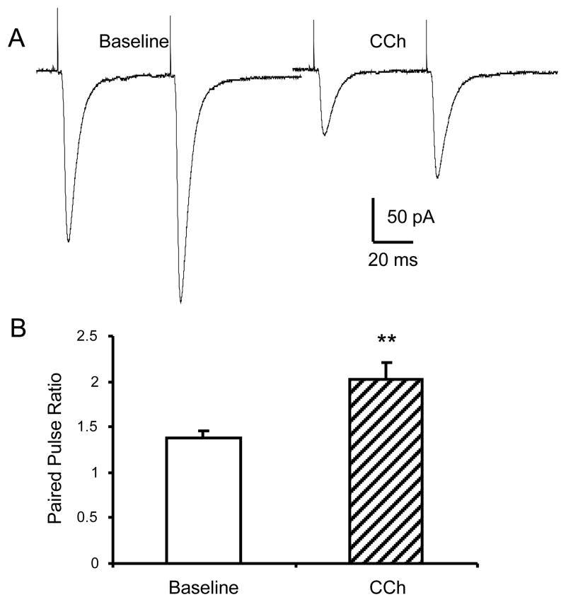
The anterolateral cell group of the bed nucleus of the stria terminalis (BNST(ALG)) serves as an important relay station in stress circuitry. Limbic inputs to the BNST(ALG) are primarily glutamatergic and activity-dependent changes in this input have been implicated in abnormal behaviors associated with chronic stress and addiction. Significantly, local infusion of acetylcholine (ACh) receptor agonists into the BNST trigger stress-like cardiovascular responses, however, little is known about the effects of these agents on glutamatergic transmission in the BNST(ALG). Here, we show that glutamate- and ACh-containing fibers are found in close association in the BNST(ALG). Moreover, in the presence of the acetylcholinesterase inhibitor, eserine, endogenous ACh release evoked a long-lasting reduction of the amplitude of stimulus-evoked EPSCs. This effect was mimicked by exogenous application of the ACh analog, carbachol, which caused a reversible, dose-dependent, reduction of the evoked EPSC amplitude, and an increase in both the paired-pulse ratio and coefficient of variation, suggesting a presynaptic site of action. Uncoupling of postsynaptic G-proteins with intracellular GDP-β-S, or application of the nicotinic receptor antagonist, tubocurarine, failed to block the carbachol effect. In contrast, the carbachol effect was blocked by prior application of atropine or M(2) receptor-preferring antagonists, and was absent in M(2)/M(4) receptor knockout mice, suggesting that presynaptic M(2) receptors mediate the effect of ACh. Immunoelectron microscopy studies further revealed the presence of M(2) receptors on axon terminals that formed asymmetric synapses with BNST neurons. Our findings suggest that presynaptic M(2) receptors might be an important modulator of the stress circuit and hence a novel target for drug development.


Figures

Fig 1
Fig 1
Close correlation of glutamate fibers and ACh fibers in the BNSTALG. Photomicrographs showing Thy-1-eYFP expression in the BLA (A) and the BNST (B) of transgenic Thy-1 mice (10x, scale bar = 100 μm, aa: anterior commissure; CP: Caudate Putamen; ec: external capsulae; ic: internal capsulae; LV: lateral ventricle; st: stria terminalis). Arrows indicate stria terminalis bundle arising from the BLA (A) and entering the BNST (B). C-C″ Dual immunofluorescence experiment demonstrated that both Thy-1-YFP-positive fibers (C, green) and choline acetyltransferase-positive (ChAT, C′, red) fibers are highly immunoreactive in the BNSTALG and they course in parallel in the dorso-ventral direction. Occasionally, Thy-1 and ChAT-positive fibers overlap and make point contacts which each other as indicated by arrows (C″, merged, 63x, scale bar 10 μm).
Fig 2
Fig 2
Endogenous ACh and the ACh analogue CCh decreased the amplitude of eEPSCs in the BNSTALG. A, upper traces showing eEPSCs recorded before (1), during (2) and after (3) eserine application. Eserine decreased the amplitude of eEPSCs, an effect that was completely blocked by prior application of atropine (5 μM). *, p<0.05 vs atropine. B, CCh (10 μM) application decreased the amplitude of eEPSCs, an effect that reversed after 10 min of wash with ACSF, **, p<0.01. C, A dose-response curve for the CCh-mediated decrease in eEPSC amplitude revealed an EC50 of 4.0 μM. The numbers of neurons tested at each CCh concentration were between 4 and 10. Scale bar: 10 ms, 100 pA.
Fig 3
Fig 3
The decrease of eEPSC amplitude during CCh application is associated with an increase of PPR. A, In the presence of CCh, an increased PPR was observed associated with the decrease of eEPSC amplitude. B, A bar chart showing group data for the mean increase in PPR during CCh application; **, paired t-test, p<0.01.
Fig 4
Fig 4
CCh decreased the frequency of mEPSCs. Spontaneous mEPSCs in BNST were recorded in the presence of TTX (1 μM) and GABAA antagonist SR 95531 (5 μM). A, Representative traces showing mEPSCs recorded from BNSTALG neurons before and during CCh (10 μM) application (right). B, Group data indicated CCh reduced the frequency but not the amplitude of mEPSCs. n=11, **, paired t-test p<0.01. C, Cumulative data plots showing that CCh increased the inter-event interval but not the amplitude of mEPSCs.
Fig 5
Fig 5
CCh has no effect on postsynaptic AMPA current induced by local pressure ejection of AMPA (1 mM). A, Representative traces showed postsynaptic currents induced by pressure application of AMPA, which were not affected by CCh application but were blocked by DNQX (20 μM). B, Group data showing that no change of postsynaptic AMPA current was observed during CCh application in the presence or absence of TTX (1 μM).
Fig 6
Fig 6
Uncoupling postsynaptic G-protein-coupled receptors with intracellular GDP-β-S had no effect on the CCH response. A, In neurons recorded with intracellular GDP-β-S, CCh reversibly decreased the amplitude of eEPSCs B, Group data showing that the CCh effect in the presence of GDP-β-S was not different from control neurons recorded with regular patch solution. C,D, Similarly, intracellular inclusion of GDP-β-S had no effect on the CCh-induced increase of PPR, which was similar to that of control neurons. *, p<0.05, **, p<0.01 vs baseline.
Fig 7
Fig 7
Activation of muscarinic receptors, but not nicotinic receptors, mediates the CCh effect. AB, Histograms showing that the effect of CCh on the eEPSC amplitude and PPR was completely blocked by the muscarinic antagonist atropine (5 μM), but not by the nicotinic antagonist tubocurarine (TC, 10 μM). C,D, Histograms showing that the muscarinic receptor agonist, muscarine, mimicked the inhibitory effect the CCh on eEPSC amplitude, and increased the PPR. E, The CCh (10 μM) effect was blocked or attenuated by M1 or M2 subtype selective antagonists, drug concentration (in μM): TZP, telenzepine 1, 10; PZP, pirenzepine 1, 10; DMTD, dimethindene 1, 10; MTC, methoctramine 1, 10; but not by M4 antagonist PD102807, 10. *, p<0.05; **, p<0.01.
Fig 8
Fig 8
CCh effects in wild type, M1−/−, and M2/M4−/− mice. A, Expression of muscarinic receptors mRNA in brain tissue from wild type (WT), M1−/−, M2/M4−/− mice. B, Representative traces showing the CCh effects on the amplitude of eEPSCs and PPR in these mouse lines. C, Group data showing that the effect of CCh on the eEPSC amplitude remained intact in M1−/− mice, but was significantly attenuated in M2/M4−/− mice (p<0.01, vs WT). D, Group data showing that the PPR was also significantly increased during CCh application in WT, M1−/− mice but not M2/M4−/− mice. *, p<0.05,** p<0.01.
Fig 9
Fig 9
Diaminobenzidine label for the M2 acetylcholine receptor (arrows) identified in axon terminals in the BNST. These terminals were frequently observed to make asymmetric synaptic contacts with dendritic spines (A and B) as well as dendritic shafts (B). Immunoreactive axon terminals were sometimes noted to contain dense core vesicles (arrowheads, B and C). Scale bar is 500 nm.

Similar articles

Cited by

References

    1. Adamec R. The relationship between the amygdala and bed nucleus of the stria terminalis in the cat: an evoked potential and single cell study. Behav Neural Biol. 1989;52:295–320. - PubMed
    1. Albuquerque EX, Pereira EF, Alkondon M, Rogers SW. Mammalian nicotinic acetylcholine receptors: from structure to function. Physiol Rev. 2009;89:73–120. - PMC - PubMed
    1. Alves FH, Crestani CC, Resstel LB, Correa FM. Cardiovascular effects of carbachol microinjected into the bed nucleus of the stria terminalis of the rat brain. Brain Res. 2007;1143:161–168. - PubMed
    1. Aoki C, Kabak S. Cholinergic terminals in the cat visual cortex: ultrastructural basis for interaction with glutamate-immunoreactive neurons and other cells. Vis Neurosci. 1992;8:177–191. - PubMed
    1. Bellingham MC, Berger AJ. Presynaptic depression of excitatory synaptic inputs to rat hypoglossal motoneurons by muscarinic M2 receptors. J Neurophysiol. 1996;76:3758–3770. - PubMed

Publication types

MeSH terms