. 2012 Feb 1;72(3):736-46.
doi: 10.1158/0008-5472.CAN-11-2584. Epub 2011 Dec 13.

A pharmacologic inhibitor of the protease Taspase1 effectively inhibits breast and brain tumor growth

Affiliations

A pharmacologic inhibitor of the protease Taspase1 effectively inhibits breast and brain tumor growth

David Y Chen et al. Cancer Res. .

Abstract

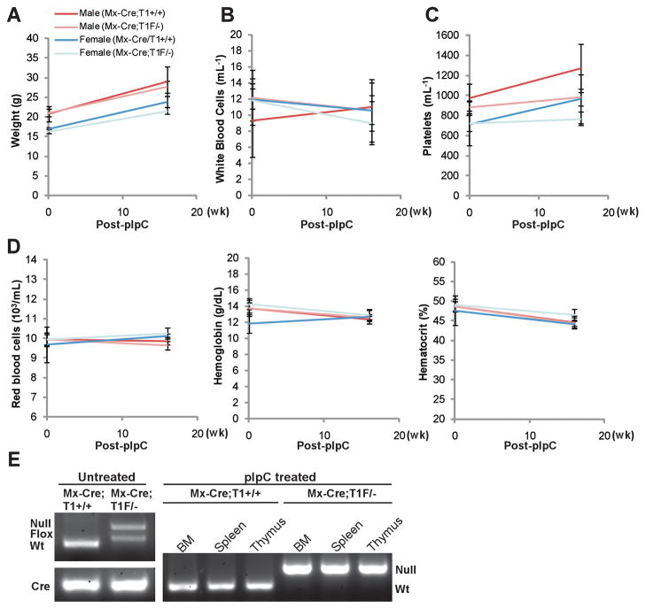
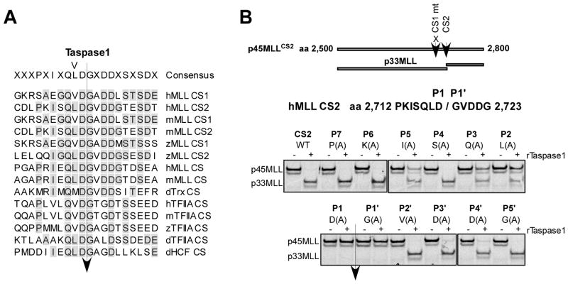
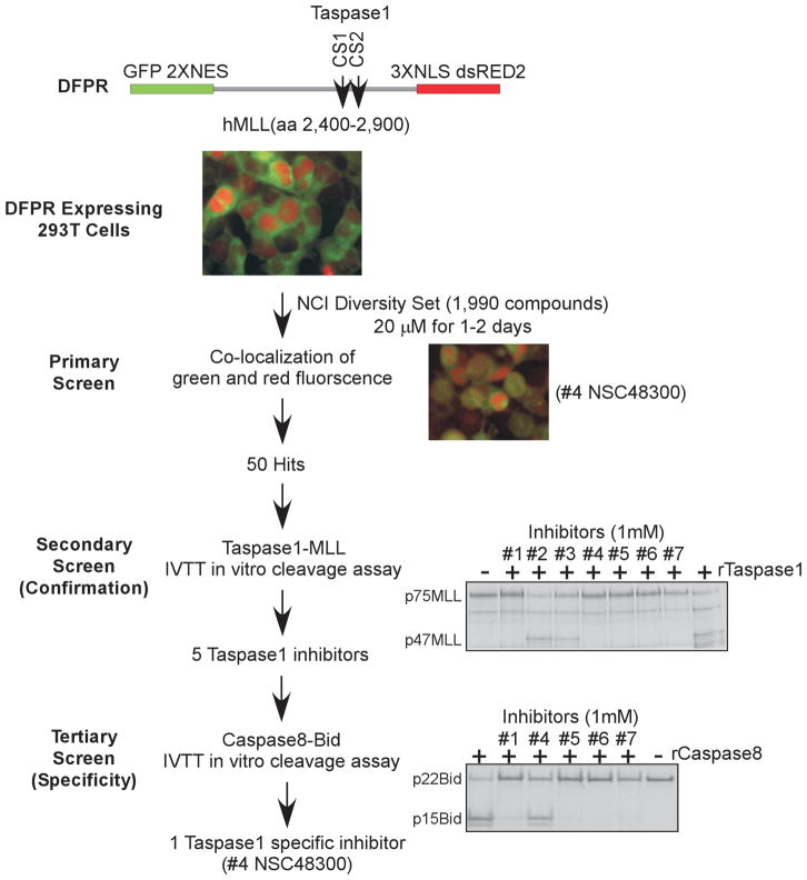
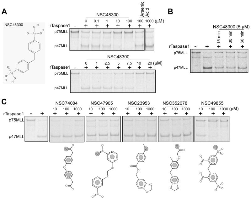
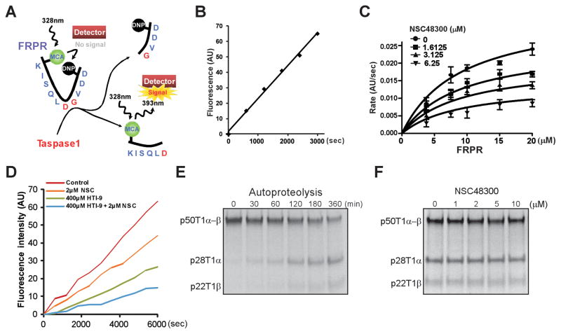
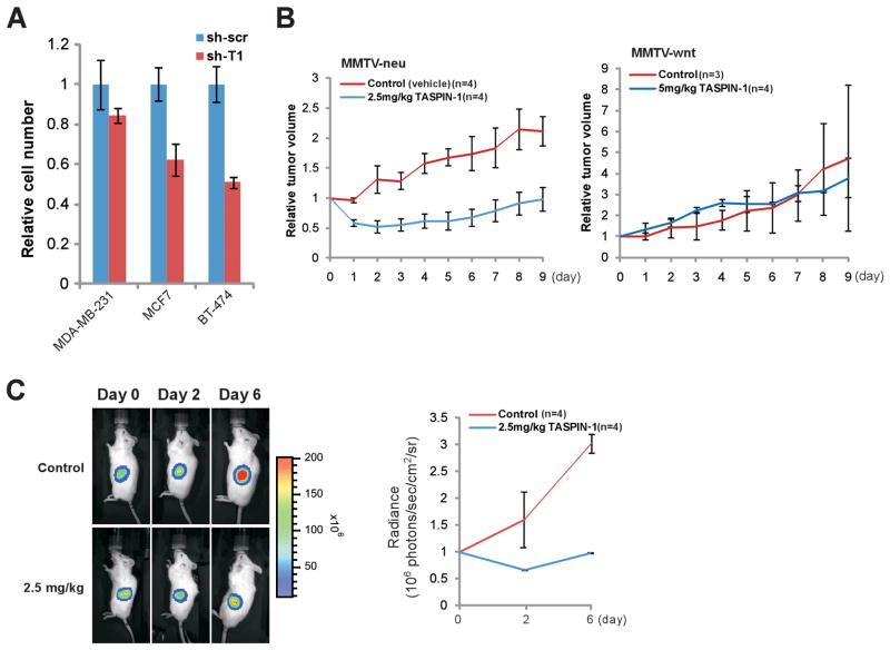
The threonine endopeptidase Taspase1 has a critical role in cancer cell proliferation and apoptosis. In this study, we developed and evaluated small molecule inhibitors of Taspase1 as a new candidate class of therapeutic modalities. Genetic deletion of Taspase1 in the mouse produced no overt deficiencies, suggesting the possibility of a wide therapeutic index for use of Taspase1 inhibitors in cancers. We defined the peptidyl motifs recognized by Taspase1 and conducted a cell-based dual-fluorescent proteolytic screen of the National Cancer Institute diversity library to identify Taspase1 inhibitors (TASPIN). On the basis of secondary and tertiary screens the 4-[(4-arsonophenyl)methyl]phenyl] arsonic acid NSC48300 was determined to be the most specific active compound. Structure-activity relationship studies indicated a crucial role for the arsenic acid moiety in mediating Taspase1 inhibition. Additional fluorescence resonance energy transfer-based kinetic analysis characterized NSC48300 as a reversible, noncompetitive inhibitor of Taspase1 (K(i) = 4.22 μmol/L). In the MMTV-neu mouse model of breast cancer and the U251 xenograft model of brain cancer, NSC48300 produced effective tumor growth inhibition. Our results offer an initial preclinical proof-of-concept to develop TASPINs for cancer therapy.


Conflict of interest statement

Disclosure of Potential Conflicts of Interest

No potential conflicts of interests were disclosed.

Figures

Figure 1
Figure 1
Induced genetic deletion of Taspase1 is well-tolerated in adult mice. A, Mice bearing an interferon-inducible cre recombinase (Mx-Cre) transgene and the indicated alleles of Taspase1 (+, wild-type; −, knockout; F, conditional allele) at 7 weeks of age were subjected to 5 doses of polyinosine-polycytidine (pIpC) injection. Baseline measurements were obtained at 6 weeks of age. Mice were sacrificed 16 weeks after the last dose of pIpC to obtain post-treatment measurements and harvest bone marrow (BM), spleen, and thymus for genotypic analysis. B, C, and D, Genetic deletion of Taspase1 dose not affect the parameters of white blood cell (B), platelet (C), and red blood cell counts (D). E, Genotyping of the indicated tissues demonstrates a complete deletion of Taspase1 after the pIpC treatment.
Figure 2
Figure 2
Characterization of the Taspase1 cleavage site consensus motif. A, Alignment of known Taspase1 cleavage sites (CS). h, m, z, and d denote human, mouse, zebra fish, and Drosophila, respectively. B, Alanine scanning mutagenesis of the CS2 region of human MLL. The indicated p45MLLCS2 (aa 2,500 to 2,800) cleavage reporters were generated by single alanine substitution of individual amino acid across P7 to P5′ of CS2, where the CS1 was invariably mutated to enable assessment of a single cleavage site. The individual IVTT, 35S-methionine labeled, p45MLLCS2 reporters were incubated with 15 ng of rTaspase1 (recombinant Taspase1) for 30 minutes at 30°C, resolved by SDS-PAGE, and monitored by autoradiography.
Figure 3
Figure 3
A Novel, cell-based, dual fluorescent Taspase1 proteolytic reporter (DFPR) identified small molecule TASPINs. Schematics depict the design of our three-tiered screen for TASPINs. In the primary screen, DFPR expressing 293T HEK cells were treated with 20 μM of the NCI DTP Diversity Set compounds for 2 days, and the fluorescence was determined at day 1 and 2. Pictures of DFPR expressing 293T HEK cells, either mock treated (DMSO) as a negative control (top middle color picture) or compound #4 NSC48300 treated as a positive illustration (lower right color picture), are presented. In the secondary (confirmation) screen, 15 ng of rTaspase1 was incubated with 1 mM of the indicated compounds for 30 minutes before adding the radiolabeled p75MLL (aa 2,400–2,900) cleavage reporter for additional 30 minutes at 30°C. In the tertiary (specificity) screen, 250 ng of rCaspase8 was incubated with 1 mM of the indicated compounds for 30 minutes before adding the radiolabeled p22Bid cleavage reporter for 2 hours at 30°C. Cleavage was assessed by SDS-PAGE and autoradiography.
Figure 4
Figure 4
Characterization of NSC48300 and determination of its structure-activity relationship. A, Chemical structure of NSC48300. 5 ng of rTaspase1 was incubated with the indicated concentrations of either NSC48300 or arsenic acid for 30 minutes at room temperature before adding the radiolabeled p75MLL reporter for 30 minutes at 30°C. B, 5 ng of rTaspase1 was incubated with 5 μM of NSC48300 for the indicated periods of time at room temperature before adding the radiolabeled p75MLL reporter for 30 minutes at 30°C. C, 5 ng of rTaspase1 was incubated with the indicated concentrations of individual NSC48300 analogues for 30 minutes at room temperature before adding the radiolabeled p75MLL reporter for 30 minutes at 30°C. Cleavage was assessed by SDS-PAGE and autoradiography.
Figure 5
Figure 5
Enzyme kinetics analyses of NSC48300. A, Diagram depicts the FRPR. B, 15 μM of FRPR was incubated with 100 nM of rTaspase1 and the fluorescence was monitored. AU denotes absolute fluorescence units. C, Non-linear regression of the reaction curves revealed that NSC48300 is a non-competitive inhibitor of Taspase1. Increasing concentrations of FRPR were incubated with 100 nM of rTaspase1 that was pretreated with the indicated concentrations of NSC48300. Data presented are mean ± SD of three independent experiments. D, 100nM of Taspase1 was pre-incubated for 30 minutes with the indicated concentrations of NSC48300 (NSC) and/or HTI-9 before the addition of FRPR. Reaction progress curves are shown. E, Precursor Taspase1 (p50T1α-β) undergoes autoproteolysis to generate 28kD α (p28T1α) and 22kD β (p22T1β) subunits. 0.5 μl of radiolabeled, 50kD precursor Taspase1 (p50T1α-β) was incubated at 30°C for the indicated periods of time. F, 0.5 μl of radiolabeled p50T1α-β was incubated at 30°C for 6 hours in the presence of indicated concentrations of NSC48300.
Figure 6
Figure 6
The cellular sensitivity to NSC48300 correlates with the protein expression of Taspase1. A, The indicated MEFs were treated with 1uM of NSC48300 for 2 days and the relative cell number was measured by MTT assays. Values obtained from the parental Taspase1−/− MEFs after NSC48300 treatment were designated as 1. Data presented are mean ± SD of three independent experiments. B, The NSC48300 GI50 (growth inhibition 50%) data were acquired through a publically available database (35). To compare relative sensitivity among different brain tumor cell lines, log(GI50) values of each individual lines were obtained. The standard deviations of individual GI50 from the mean were plotted (positive values indicate higher sensitivity). The protein levels of Taspase1 in the indicated cell lines have been reported and are represented as a bar graph. The indicated brain cancer cell lines were treated with increasing concentration of NSC48300 for 2 days plus or minus 100nM LPA and then subjected to MTT assays. Data presented are mean ± SD of two triplicate independent experiments. C, A positive correlation of Taspase1 protein levels and cellular sensitivity to NSC48300 in the NCI60 breast tumor cell lines. Data collection, experimentations, and analyses were performed as described in B on the indicated breast cancer cell lines.
Figure 7
Figure 7
NSC48300 disrupts breast and brain cancer growth in mouse cancer models. A, The indicated human breast cancer cell lines with control (sh-scr) or Taspase1 (sh-T1) knockdown were plated, and cell numbers were counted 4 days after the initial plating. The average cell number of control knockdown cells was assigned as 1. Data presented are mean ± SD of three independent experiments. B, Tumor bearing MMTV-neu or MMTV-wnt mice were treated at day 0 when de novo tumors reached 1cm at the longest dimension. NSC48300 was given at the indicated doses every other day for a total of 5 intravenous injections. Graphs represent the relative tumor volume throughout the course of treatment. The tumor size measured at day 0 immediately before treatment was assigned as 1. Data presented are mean ± SD of the indicated number of tumors of each arm. C, U251 cells transduced with a luciferase reporter were implanted into the flanks of male NOD-scid IL2Rγ−/− mice at 6–8 weeks of age (day −1). Mice were then treated on days 0, 2, and 4 with the indicated dose of NSC48300. Tumor mass was determined by bioluminescent imaging using an IVIS 100 and representative bioluminescent images are presented. The graph represents mean ± SD of 4 tumors from each arm. The absolute bioluminescence was normalized to a value of 1 at day 0.

Comment in

  • Targeting Taspase1 for cancer therapy--letter.
    Stauber RH, Bier C, Knauer SK. Stauber RH, et al. Cancer Res. 2012 Jun 1;72(11):2912; author reply 2913. doi: 10.1158/0008-5472.CAN-12-0150. Epub 2012 May 16. Cancer Res. 2012. PMID: 22593195 No abstract available.

Similar articles

Cited by

References

    1. Furie B, Furie BC. Molecular and cellular biology of blood coagulation. N Engl J Med. 1992;326:800–6. - PubMed
    1. Zaman MA, Oparil S, Calhoun DA. Drugs targeting the renin-angiotensin-aldosterone system. Nat Rev Drug Discov. 2002;1:621–36. - PubMed
    1. Brown MS, Ye J, Rawson RB, Goldstein JL. Regulated intramembrane proteolysis: a control mechanism conserved from bacteria to humans. Cell. 2000;100:391–8. - PubMed
    1. Ye Y, Fortini ME. Proteolysis and developmental signal transduction. Semin Cell Dev Biol. 2000;11:211–21. - PubMed
    1. Thornberry NA, Lazebnik Y. Caspases: enemies within. Science. 1998;281:1312–6. - PubMed

Publication types

MeSH terms