Review
. 2012 Jan 1;4(1):a008821.
doi: 10.1101/cshperspect.a008821.

Autophagy and cancer

Affiliations
Review

Autophagy and cancer

Li Yen Mah et al. Cold Spring Harb Perspect Biol. .

Abstract

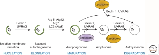
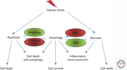
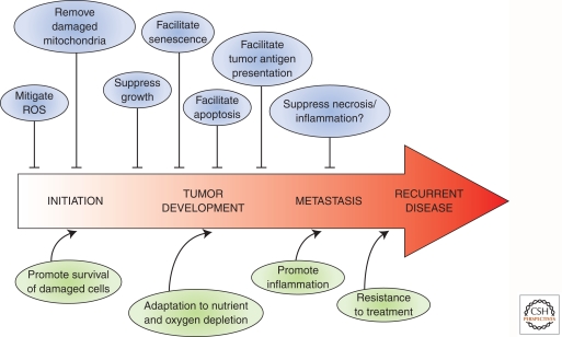
(Macro)autophagy is a cellular membrane trafficking process that serves to deliver cytoplasmic constituents to lysosomes for degradation. At basal levels, it is critical for maintaining cytoplasmic as well as genomic integrity and is therefore key to maintaining cellular homeostasis. Autophagy is also highly adaptable and can be modified to digest specific cargoes to bring about selective effects in response to numerous forms of intracellular and extracellular stress. It is not a surprise, therefore, that autophagy has a fundamental role in cancer and that perturbations in autophagy can contribute to malignant disease. We review here the roles of autophagy in various aspects of tumor suppression including the response of cells to nutrient and hypoxic stress, the control of programmed cell death, and the connection to tumor-associated immune responses.


Figures

Figure 1.
Figure 1.
Cellular mechanism and molecular regulators of autophagy in eukaryotes. NUCLEATION: Beclin 1(Atg6) and UVRAG (UV irradiation resistance–associated gene), are required for the formation of the isolation membrane for sequestering the autophagic substrate. ELONGATION: The closure of the isolation membrane to form autophagosomes causes sequestration/entrapment of cytoplasmic constituents. This requires the conjugation of Atg5 and Atg12 and is catalyzed by Atg7 (E1-like enzyme). Meanwhile, pro-LC3 (Atg8) is cleaved to form LC3-I, which is then lipidated to form LC3-II. MATURATION: Autophagosomes dock and fuse with lysosomes to form autolysosomes. Although the molecular mechanism is not fully understood, Beclin 1 and UVRAG are thought to mediate this process. Alternatively, autophagosomes can fuse with endosomal vesicles, such as endosomes and multivesicular bodies, to form amphisomes, which eventually dock with lysosomes. DEGRADATION: After fusion with lysosomes, autolysosomes are generated where the sequestered materials are hydrolyzed. The inner membrane of autophagosomes and the cargoes are degraded by lysosomal enzymes, and breakdown products are released back into the cytosol.
Figure 2.
Figure 2.
p53 modulates autophagy in multiple ways. Basal levels of p53 target repression of autophagy from within the cytoplasm. In response to cellular stress, the levels of p53 become elevated and accumulate in the nucleus. This results in activation of a series of target genes that positively regulate autophagy. PUMA and BAX also localize to mitochondria. DRAM-1, in contrast, localizes to lysosomes, and Sestrin-2 modulates autophagy via mTOR. SESN2, the gene encoding Sestrin-2.
Figure 3.
Figure 3.
Autophagy and the control of cell death. There is cross talk between apoptosis and autophagy in the control of cell death. The two pathways also repress one another, fighting for either cell survival or cell death. Autophagy also regulates necrosis. Both pathways can regulate each other, and the products of inflammation can positively feed back to enhance the amplitude of these responses. Examples are shown to indicate how apoptosis and autophagy and necrosis and autophagy are connected.
Figure 4.
Figure 4.
The contrasting roles of autophagy in cancer. Activating autophagy at different stages of cancer yields multiple opposing effects. In healthy cells, autophagy prevents cellular transformation by removing ROS and damaged mitochondria. However, following transformation, activation of autophagy can promote and suppress cancer progression, depending on the timing or stage of disease. Autophagy either mediates its effects directly or “communicates” with other cellular pathways such as senescence, apoptosis, necrosis, and inflammation.

Similar articles

Cited by

References

    1. Adams PD 2009. Healing and hurting: Molecular mechanisms, functions, and pathologies of cellular senescence. Mol Cell 36: 2–14 - PubMed
    1. Aita VM, Liang XH, Murty VV, Pincus DL, Yu W, Cayanis E, Kalachikov S, Gilliam TC, Levine B 1999. Cloning and genomic organization of beclin 1, a candidate tumor suppressor gene on chromosome 17q21. Genomics 59: 59–65 - PubMed
    1. Aki T, Yamaguchi K, Fujimiya T, Mizukami Y 2003. Phosphoinositide 3-kinase accelerates autophagic cell death during glucose deprivation in the rat cardiomyocyte-derived cell line H9c2. Oncogene 22: 8529–8535 - PubMed
    1. Amaravadi RK, Thompson CB 2007. The roles of therapy-induced autophagy and necrosis in cancer treatment. Clin Cancer Res 13: 7271–7279 - PubMed
    1. Balkwill F, Charles KA, Mantovani A 2005. Smoldering and polarized inflammation in the initiation and promotion of malignant disease. Cancer Cell 7: 211–217 - PubMed

Publication types

LinkOut - more resources