

Room-temperature susceptometry predicts biopsy-determined hepatic iron in patients with elevated serum ferritin
- PMID: 22166564
- PMCID: PMC3499026
Room-temperature susceptometry predicts biopsy-determined hepatic iron in patients with elevated serum ferritin
Abstract
Background: There is an ongoing clinical need for novel methods to measure hepatic iron content (HIC) noninvasively. Both magnetic resonance imaging (MRI) and superconducting quantum interference device (SQUID) methods have previously shown promise for estimation of HIC, but these methods can be expensive and are not widely available. Room-temperature susceptometry (RTS) represents an inexpensive alternative and was previously found to be strongly correlated with HIC estimated by SQUID measurements among patients with transfusional iron overload related to thalassemia.
Aim: The goal of the current study was to examine the relationship between RTS and biochemical HIC measured in liver biopsy specimens in a more varied patient cohort.
Material and methods: Susceptometry was performed in a diverse group of patients with hyperferritinemia due to hereditary hemochromatosis (HHC) (n = 2), secondary iron overload (n = 3), nonalcoholic fatty liver disease (NAFLD) (n = 2), and chronic viral hepatitis (n = 3) within one month of liver biopsy in the absence of iron depletion therapy.
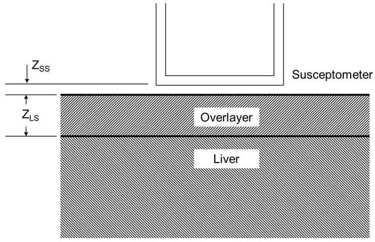
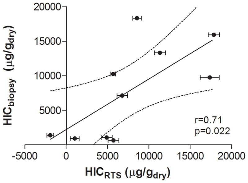
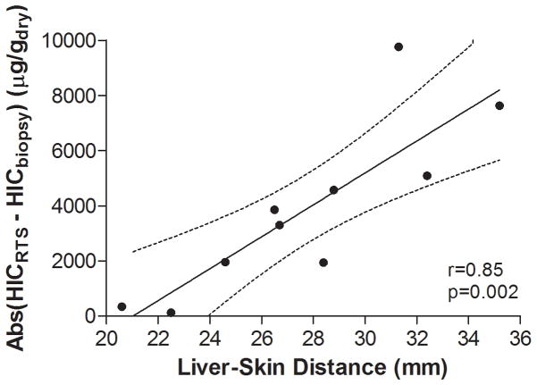
Results: The correlation coefficient between HIC estimated by susceptometry and by biochemical iron measurement in liver tissue was 0.71 (p = 0.022). Variance between liver iron measurement and susceptometry measurement was primarily related to reliance on the patient's body-mass index (BMI) to estimate the magnetic susceptibility of tissue overlying the liver.
Conclusions: We believe RTS holds promise for noninvasive measurement of HIC. Improved measurement techniques, including more accurate overlayer correction, may further improve the accuracy of liver susceptometry in patients with liver disease.
Figures
Similar articles
-
Susceptibility based multiparametric quantification of liver disease: Non-invasive evaluation of steatosis and iron overload.Magn Reson Imaging. 2019 Nov;63:114-122. doi: 10.1016/j.mri.2019.08.016. Epub 2019 Aug 16. Magn Reson Imaging. 2019. PMID: 31425813
-
Measurement of liver iron concentration by superconducting quantum interference device biomagnetic liver susceptometry validates serum ferritin as prognostic parameter for allogeneic stem cell transplantation.Eur J Haematol. 2016 Oct;97(4):336-41. doi: 10.1111/ejh.12734. Epub 2016 Feb 29. Eur J Haematol. 2016. PMID: 26800433
-
Hepcidin levels correlate to liver iron content, but not steatohepatitis, in non-alcoholic fatty liver disease.BMC Gastroenterol. 2018 Jun 5;18(1):78. doi: 10.1186/s12876-018-0804-0. BMC Gastroenterol. 2018. PMID: 29871592 Free PMC article.
-
Diagnostic and therapeutic implications of the association between ferritin level and severity of nonalcoholic fatty liver disease.World J Gastroenterol. 2012 Aug 7;18(29):3782-6. doi: 10.3748/wjg.v18.i29.3782. World J Gastroenterol. 2012. PMID: 22876027 Free PMC article. Review.
-
A diagnostic approach to hyperferritinemia with a non-elevated transferrin saturation.J Hepatol. 2011 Aug;55(2):453-8. doi: 10.1016/j.jhep.2011.02.010. Epub 2011 Feb 24. J Hepatol. 2011. PMID: 21354228 Review.
Cited by
-
Serum or plasma ferritin concentration as an index of iron deficiency and overload.Cochrane Database Syst Rev. 2021 May 24;5(5):CD011817. doi: 10.1002/14651858.CD011817.pub2. Cochrane Database Syst Rev. 2021. PMID: 34028001 Free PMC article.
-
Non-invasive diagnosis of alcoholic liver disease.World J Gastroenterol. 2014 Oct 28;20(40):14626-41. doi: 10.3748/wjg.v20.i40.14626. World J Gastroenterol. 2014. PMID: 25356026 Free PMC article. Review.
-
A Novel Magnetoelectric Biomagnetic Susceptometer on Iron Level Detection with Mice Tissue in Vitro.Med Devices Sens. 2018 Feb;1(1):e10004. doi: 10.1002/mds3.10004. Epub 2018 Jun 19. Med Devices Sens. 2018. PMID: 30643870 Free PMC article.
References
-
- Bonkovsky HL, Lambrecht RW, Shan Y. Iron as a co-morbid factor in nonhemochromatotic liver disease. Alcohol. 2003;30:137–144. - PubMed
-
- Zamcheck N, Klasusenstock O. Liver biopsy. II. The risk of needle biopsy. N Engl J Med. 1953;249:1062–1069. - PubMed
-
- Butensky E, Fischer R, Hudes M, Schumacher L, Williams R, Moyer TP, Vichinsky E, Harmatz P. Variability in hepatic iron concentration in percutaneous needle biopsy specimens from patients with transfusional hemosiderosis. Am J Clin Pathol. 2005;123:146–152. - PubMed
-
- Nielsen P, Gunther U, Durken M, Fischer R, Dullmann J. Serum ferritin iron in iron overload and liver damage: correlation to body iron stores and diagnostic relevance. J Lab Clin Med. 2000;135:413–418. - PubMed
Publication types
MeSH terms
Substances
Grants and funding
LinkOut - more resources
Full Text Sources
Medical
