Review
. 2012 Feb;18(1):68-80.
doi: 10.1017/S1431927611012402. Epub 2011 Dec 14.

Endothelial cell-pericyte interactions stimulate basement membrane matrix assembly: influence on vascular tube remodeling, maturation, and stabilization

Affiliations
Review

Endothelial cell-pericyte interactions stimulate basement membrane matrix assembly: influence on vascular tube remodeling, maturation, and stabilization

Amber N Stratman et al. Microsc Microanal. 2012 Feb.

Abstract

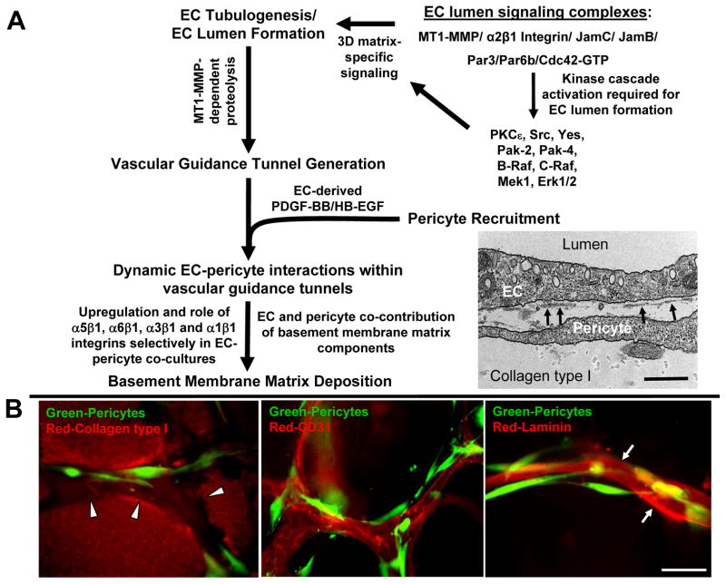
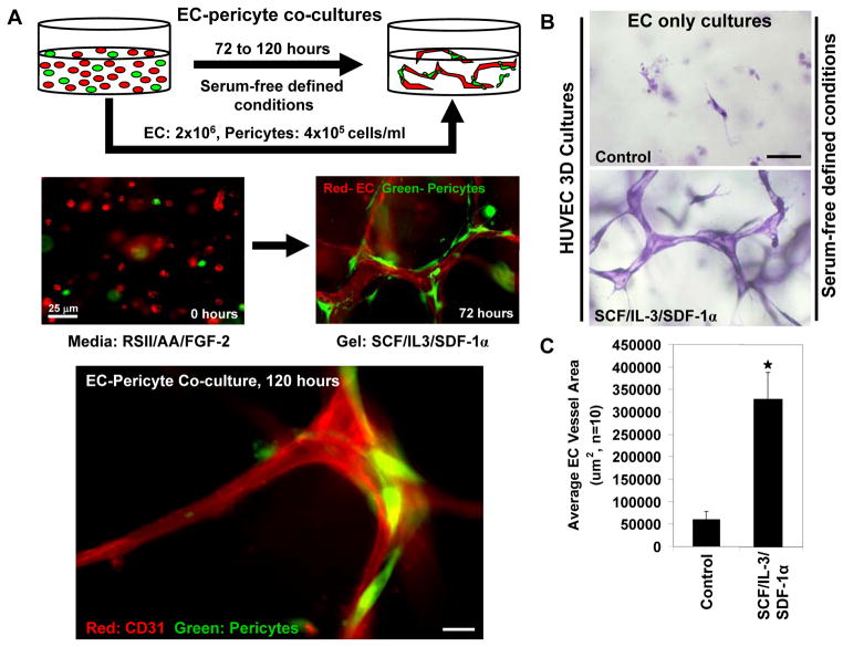
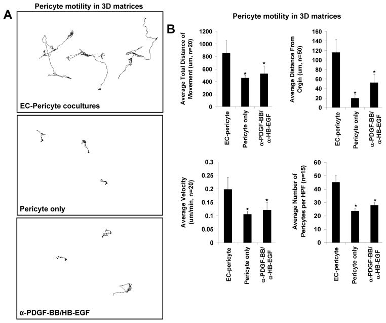
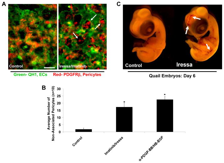
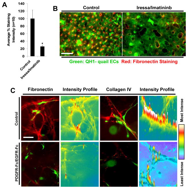
Extracellular matrix synthesis and deposition surrounding the developing vasculature are critical for vessel remodeling and maturation events. Although the basement membrane is an integral structure underlying endothelial cells (ECs), few studies, until recently, have been performed to understand its formation in this context. In this review article, we highlight new data demonstrating a corequirement for ECs and pericytes to properly deposit and assemble vascular basement membranes during morphogenic events. In EC only cultures or under conditions whereby pericyte recruitment is blocked, there is a lack of basement membrane assembly, decreased vessel stability (with increased susceptibility to pro-regressive stimuli), and increased EC tube widths (a marker of dysfunctional EC-pericyte interactions). ECs and pericytes both contribute basement membrane components and, furthermore, both cells induce the expression of particular components as well as integrins that recognize them. The EC-derived factors--platelet derived growth factor-BB and heparin binding-epidermal growth factor--are both critical for pericyte recruitment to EC tubes and concomitant vascular basement membrane formation in vitro and in vivo. Thus, heterotypic EC-pericyte interactions play a fundamental role in vascular basement membrane matrix deposition, a critical tube maturation event that is altered in key disease states such as diabetes and cancer.


Figures

Figure 1
Figure 1. Molecular mechanisms controlling EC tubulogenesis, pericyte recruitment and tube stabilization in 3D matrices
(A) The schematic depicts the role of MT1-MMP proteolysis, EC lumen signaling complexes, and signal transduction cascades on the promotion of early EC morphogenic events including lumenogenesis and vascular guidance tunnel formation. Pericyte recruitment occurs to these developing tubes through EC-derived PDGF-BB and HB-EGF leading to vascular basement membrane deposition that affects tube maturation and stabilization. EC-pericyte interactions selectively lead to increased basement membrane protein synthesis, deposition and upregulation of integrins that recognize this newly deposited ECM. The image is an electron micrograph showing EC-pericyte tube coassembly and developing vascular basement membranes depositing (arrows) between the two cell types in 3D matrices. Bar equals 0.5 μm. (B) ECs and pericytes were seeded in collagen type I matrix and allowed to assemble over 5 days. Images shown depict varying steps within this assembly process: (left) the generation and presence of vascular guidance tunnels (red: collagen type I matrix, green: GFP-pericytes); (center) the recruitment of pericytes to EC lined tubes (red: CD31, green: GFP-pericytes); (right) the deposition of the vascular basement membrane (red: laminin, green: GFP-pericytes). Bar equals 25 μm.
Figure 2
Figure 2. The development of in vitro models of EC tubulogenesis and EC-pericyte tube coassembly under defined serum-free conditions
(A) Models of EC-pericyte coassembly were developed in collagen type I matrices in which the pericytes recruit to developing EC tubes over a period of three to five days. The two cells are seeded randomly as individual cells and over time the ECs vacuolate, lumenize and interconnect into vascular networks; pericytes then recruit to these vessels to promote maturation and long term stabilization events. Images are shown to display this assembly process with the ECs immunolabeled with anti-CD31 antibodies in red and pericytes stably expressing GFP. Bar equals 10 μm. (B/C) The 3D assay systems utilized are performed using a series of three hematopoietic cytokines, SCF, SDF-1α and IL-3, to promote tubulogenesis. Images and quantification of the morphogenic phenotype observed at day 3 of culture are shown. Bar equals 25 μm.
Figure 3
Figure 3. EC-pericyte interactions promote tube remodeling, maturation and stabilization through deposition of the vascular basement membrane
EC only versus EC-pericyte cocultures were established for a period of 5 days in 3D collagen matrices. Immunostaining analysis was performed, using detergent free staining protocols to recognize only extracellular proteins, of the EC only versus EC-pericyte cocultures. Vascular basement membrane matrix assembly reveals extracellular deposition of the indicated basement membrane proteins only in the coculture setting. Pericytes are GFP labeled while the varying basement membrane proteins indicated are immunolabeled in red. Bar equals 25 μm.
Figure 4
Figure 4. EC-derived PDGF-BB and HB-EGF are required to induce pericyte motility and proliferation during EC-pericyte tube coassembly in 3D matrices
(A) Nuclear GFP-pericytes were seeded in 3D collagen matrices in the presence or absence of ECs and their motility tracked over a period of 3 days. Images of nuclear tracking of pericytes reveal the requirement of ECs to promote pericyte motility. Further, inhibition of PDGF-BB and HB-EGF in combination leads to marked suppression of pericyte motility in 3D matrices. (B) Quantification of nuclear tracking of pericytes in the presence of EC, in the absence of EC or in EC-pericyte cocultures in which PDGF-BB and HB-EGF are inhibited demonstrating that PDGF-BB and HB-EGF in combination are required to stimulate EC motility and proliferation in 3D collagen matrices.
Figure 5
Figure 5. PDGF-BB and HB-EGF direct pericyte recruitment to endothelial cell tubes during quail vascular morphogenesis
(A) Images of ECs (QH1-green) overlaid with the associated pericyte field (PDGFRβ-red) are shown demonstrating the lack of pericytes associated with EC tubes in conditions where quail embryos were treated with chemical inhibitors to PDGFRβ (Imatinib) and EGFR (Iressa) (both added at 100 nM). Bar equals 15 μm. (B) Quantification of the number of non-associated pericytes in control versus PDGF-BB/HB-EGF double inhibited treatment conditions (both antibodies added at 50 μg/ml). At day 6 of development, treatments that inhibit PDGF-BB and HB-EGF activity on pericytes lead to an increase in the number of non-associated pericytes versus controls. (C) Images of control versus treatment embryos reveals the increased incidence of cranial vascular hemorrhage phenotypes in the embryos in which pericyte recruitment is impaired.
Figure 6
Figure 6. EC-pericyte interactions markedly stimulate basement membrane deposition and assembly in 3D matrices in vitro and in vivo
(A) Quantification of fibronectin immunostaining intensity levels reveals that in conditions with disrupted EC-pericyte interactions (i.e. Imatinib/Iressa) there is a dramatic decrease in the amount of fibronectin deposition around developing quail EC-lined tubes at embryonic day 6. (B) Images of this phenotype are shown in overlay images of the vasculature (QH1-green) versus fibronectin deposition (red) in control versus treatment conditions in which pericyte recruitment is markedly inhibited. Bar equals 25 μm. (C) Images displaying collagen IV or fibronectin staining (red) versus GFP-pericytes reveals a lack of deposition in the treatment condition when pericyte recruitment is blocked. Intensity mapping of this phenotype is also included demonstrating the increased localization of deposited collagen IV around EC tubes in the control versus treated condition.

References

    1. Abramsson A, Lindblom P, Betsholtz C. Endothelial and nonendothelial sources of PDGF-B regulate pericyte recruitment and influence vascular pattern formation in tumors. J Clin Invest. 2003;112(8):1142–51. - PMC - PubMed
    1. Adams RH, Alitalo K. Molecular regulation of angiogenesis and lymphangiogenesis. Nat Rev Mol Cell Biol. 2007;8(6):464–78. - PubMed
    1. Arroyo AG, Iruela-Arispe ML. Extracellular matrix, inflammation, and the angiogenic response. Cardiovasc Res. 2010;86(2):226–35. - PMC - PubMed
    1. Astrof S, Crowley D, Hynes RO. Multiple cardiovascular defects caused by the absence of alternatively spliced segments of fibronectin. Dev Biol. 2007;311(1):11–24. - PMC - PubMed
    1. Astrof S, Hynes RO. Fibronectins in vascular morphogenesis. Angiogenesis. 2009;12(2):165–75. - PMC - PubMed

Publication types