. 2012 Feb;54(2):186-92.
doi: 10.1097/MPG.0b013e318244148b.

MicroRNA profiling identifies miR-29 as a regulator of disease-associated pathways in experimental biliary atresia

Affiliations

MicroRNA profiling identifies miR-29 as a regulator of disease-associated pathways in experimental biliary atresia

Nicholas J Hand et al. J Pediatr Gastroenterol Nutr. 2012 Feb.

Abstract

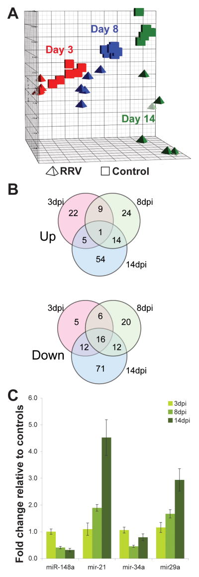
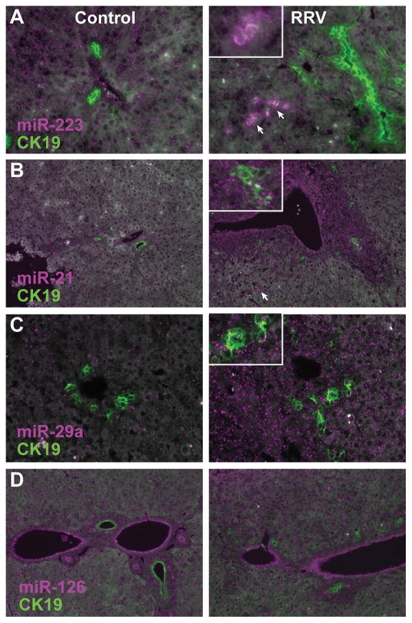
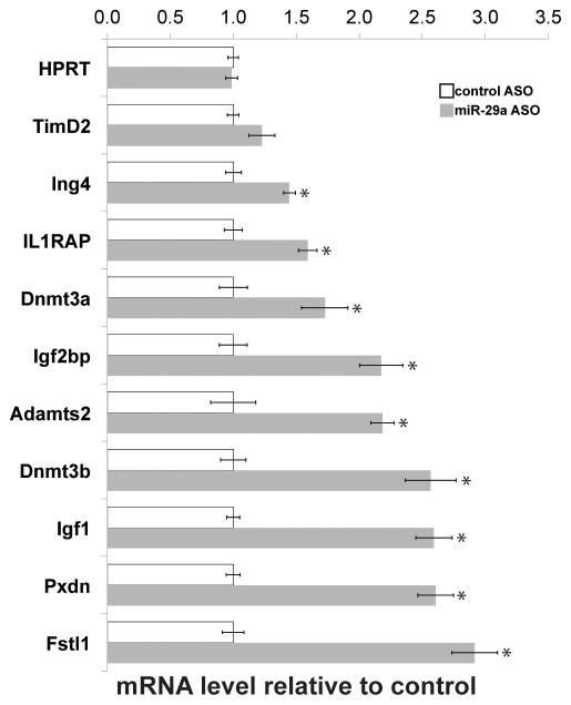
Biliary atresia (BA) is a pediatric liver disease of unknown underlying etiology, in which fibroinflammatory destruction of the extrahepatic biliary system leads to obstructive cholestasis. MicroRNAs are a class of short (18-23 nucleotide), noncoding RNA molecules, which act as negative regulators of target mRNA stability and translation. The importance of these molecules in normal and diseased liver has been demonstrated, but their potential role in the pathogenesis of BA has not been addressed. We have profiled changes in liver microRNA levels in an established mouse model of the disease, identified significantly altered transcripts, and defined the spatial expression patterns of selected microRNAs. Two of these, miR-29a/29b1, are upregulated in experimental BA. Using antisense oligonucleotide-mediated inhibition in mice, we have delineated the full set of hepatic genes regulated by miR-29 and identified 2 mRNA targets of potential pathological relevance in experimental BA, Igf1 and Il1RAP. We have used reporter assays to confirm that Igf1 and Il1RAP are direct targets of miR-29.


Figures

Figure 1
Figure 1
MicroRNA microarray summary and validation. (A) Principal component analysis of miRNA microarray data. Samples from saline-injected mice, cubes; RRV-injected, tetrahedra. Red, blue, and green represent 3dpi, 8dpi and 14dpi time points respectively. (B) Number of significantly increased and decreased miRNAs at each time point. See Supplemental Table 1 for listing of miRNAs in Fig 1B. (C) Confirmatory qPCR of selected miRNAs at the indicated time points.
Figure 2
Figure 2
In situ hybridization of (A) miR-223, (B) miR-21, (C) miR-29a, and (D) miR-126 (magenta), with simultaneous immunofluorescent detection of CK19 to detect cholangiocytes (green). In (A), the inset shows a higher magnification view of infiltrating monocytes (arrows). In (B) and (C), the insets include higher magnification views of bile ductules. Liver sections from the 8 days post saline (control) or RRV injection were hybridized with locked nucleic acid miRNA probes and a rabbit polyclonal CK19 antiserum.
Figure 3
Figure 3
In vivo inhibition of miR-29a: gene expression microarray validation. Relative transcript abundance, normalized to 28S rRNA transcript levels, of genes deregulated in response to miR-29a antisense injection, assayed by RT-qPCR of liver RNA (n=4 per group). *p<0.05.
Figure 4
Figure 4
Dual luciferase reporter assays: Igf1 and Il1RAP are direct targets of miR-29a. Igf1 and Il1RAP expression in RRV versus saline. (A) Luciferase reporter assays to detect repression of Igf1 and Il1RAP 3′ UTR sequences containing candidate miR-29a target sites by exogenous miR-29a. Results are expressed relative to a control reporter without additional UTR sequences. (B) Luciferase reporter assays to detect de-repression of Igf1 and Il1RAP 3′ UTR sequences containing candidate miR-29a target sites by a locked nucleic acid antisense oligonucleotide-mediated miR-29a inhibition. Results are expressed relative to a control oligonucleotide. (C) Relative abundance of Igf1 and Il1RAP in RRV infected mice, relative to saline controls, assayed by RT-qPCR on liver RNA at the indicated time points (n=4 per group). *p<0.05.

References

    1. Bezerra JA. Potential etiologies of biliary atresia. Pediatr Transplant. 2005;9:646–651. - PubMed
    1. Hartley JL, Davenport M, Kelly DA. Biliary atresia. Lancet. 2009;374:1704–1713. - PubMed
    1. Schreiber RA, Kleinman RE. Biliary atresia. J Pediatr Gastroenterol Nutr. 2002;35(Suppl 1):S11–16. - PubMed
    1. Mieli-Vergani G, Vergani D. Biliary atresia. Semin Immunopathol. 2009;31:371–381. - PubMed
    1. Sokol RJ, Shepherd RW, Superina R, et al. Screening and outcomes in biliary atresia: summary of a National Institutes of Health workshop. Hepatology. 2007;46:566–581. - PMC - PubMed

Publication types

MeSH terms