. 2012 Jan 25;53(1):406-12.
doi: 10.1167/iovs.11-8419.

Neuroprotective effects of recombinant T-cell receptor ligand in autoimmune optic neuritis in HLA-DR2 mice

Affiliations

Neuroprotective effects of recombinant T-cell receptor ligand in autoimmune optic neuritis in HLA-DR2 mice

Grazyna Adamus et al. Invest Ophthalmol Vis Sci. .

Abstract

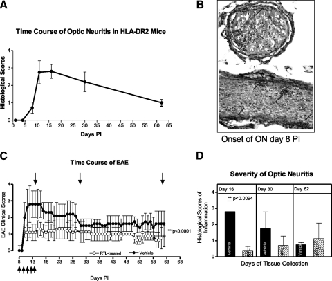
Purpose: Optic neuritis (ON) is a condition involving primary inflammation, demyelination, and axonal injury in the optic nerve and leads to apoptotic retinal ganglion cell (RGC) death, which contributes to the persistence of visual loss. Currently, ON has no effective treatment. The goal was to determine the effectiveness of immunotherapy with recombinant T-cell receptor ligand (RTL) in preventing ON in humanized HLA-DR2 transgenic mice.

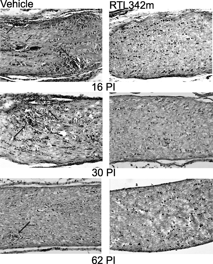
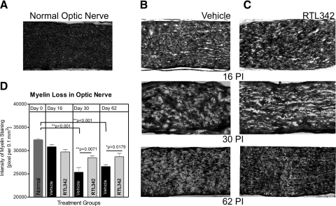
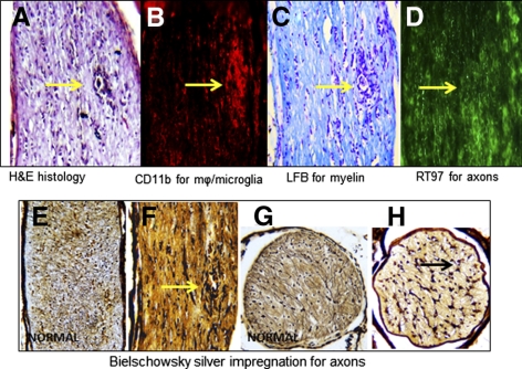
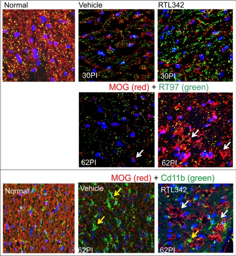
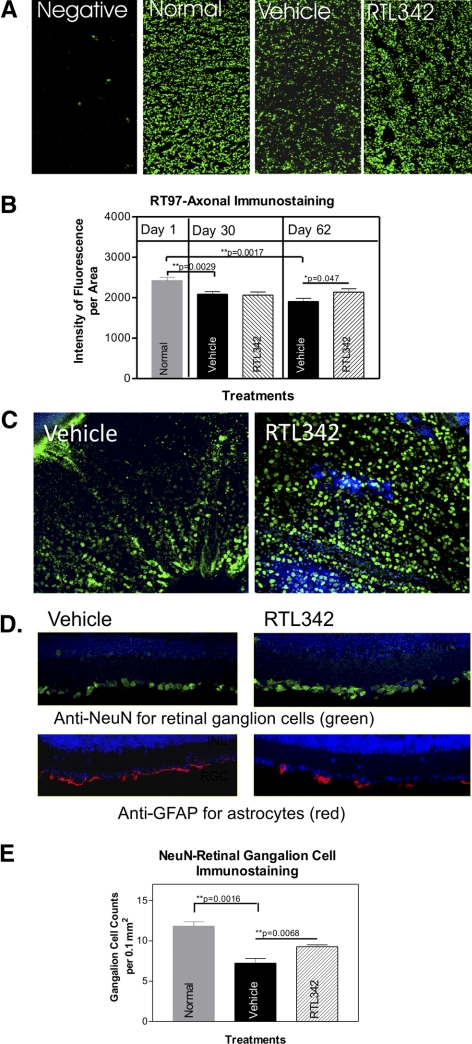
Methods: Experimental autoimmune encephalomyelitis (EAE) was induced with myelin oligodendrocyte glycoprotein in humanized HLA-DR2 (DRβ1*1501) transgenic mice. Five consecutive doses of RTL342M were administrated at the onset of ON. The development of autoimmune ON was assessed by histopathology at different time points. The levels of myelin loss, axonal loss, and RGC damage were examined by immunofluorescence.

Results: HLA-DR2 mice developed chronic ON 2 days before EAE characterized by progressive neurodegeneration in both organs. RTL342M significantly suppressed inflammation in the optic nerve and spinal cord and provided protection for at least 30 days. Examination of myelin loss showed a marked suppression of demyelination and an increase in myelin recovery in the optic nerve. Moreover, RTL342M treatment revealed a neuroprotective effect on optic nerve axons and RGCs in retinas at postimmunization (PI) day 62.

Conclusions: RTL342M suppressed clinical and histologic signs of EAE/ON by preventing the recruitment of inflammatory cells into the optic nerve and showed neuroprotective effects against ON. However, to achieve full therapeutic benefit, more doses may be needed. These findings suggest a possible clinical application of this novel class of T-cell-tolerizing drugs for patients with optic neuritis.


Figures

Figure 1.
Figure 1.
Pathology of ON associated with EAE in HLA-DR2 mice induced with MOG35-55 peptide. (A) Time course of ON in HLA-DR2 mice based on three experiments determined using longitudinal sections of the optic nerve stained with H&E. (B) Histopathology of representative ON at onset. H&E staining of the cross- and longitudinal sections; arrows: inflammatory cells. (C) Clinical time course of EAE in vehicle- and RTL342M-treated HLA-DR2 mice. Top arrows: the time of tissue collection; bottom arrows: the time of treatment administration (five doses of 20 μg RTL342M/dose on consecutive days starting at onset of ON). (D) Severity of ON in vehicle- and RTL342M-treated mice.
Figure 2.
Figure 2.
Histopathologic micrographs of HLA-DR2 mouse optic nerves collected on days 16, 30, and 62 PI. Long sections of optic nerves were stained with H&E. Arrows: inflammatory lesions.
Figure 3.
Figure 3.
Analysis of myelin loss in optic nerves of RTL342M- and vehicle-treated HLA-DR2 mice. (AC) Representative images of long optic nerve sections at ON collected on days 16, 30, and 62 PI stained with Luxol fast blue to label myelin. (A) Normal optic nerve shows compact myelin staining; longitudinal sections of optic nerves from (B) vehicle- and (C) RTL342M-treated mice collected at different times after immunization with MOG. (D) Evaluation of demyelination of optic nerves by quantification of the intensity of the staining and adjusted to 0.1 mm2 showed significantly greater demyelination in vehicle-treated mice. Values are expressed as the mean ± SEM of staining intensity from four optic nerves per group.
Figure 4.
Figure 4.
Early demyelination of optic nerves and axonal degeneration in MOG-induced ON in HLA-DR2 transgenic mice at day 16 PI (peak). Representative sequential long sections of the same optic nerve with ON stained for inflammatory cells with H&E. (A) Myelin staining with H&E; (B) macrophages (mϕ)/microglia immunofluorescent staining with anti-CD11b antibodies (red); (C) myelin staining with Luxol fast blue with cresyl blue to label cells; and (D) immunofluorescent axonal labeling using RT97 antibodies (green). (AD, arrows) An inflammatory lesion; (E–H) Bielschowsky silver impregnation for axonal staining; (E, G) normal, untreated optic nerves are populated by axons; (F, H, arrows) damaged axons.
Figure 5.
Figure 5.
Oligodendrocyte immunofluorescent labeling of the vehicle- and RTL342M-treated optic nerves. (A) Double-immunofluorescent labeling in the optic nerves collected at 30 and 62 PI with anti-MOG antibodies as a marker for oligodendrocytes and with anti-RT97 for labeling axons: anti-MOG (red), anti-RT97 (green) antibodies and colocalization (yellow). (A, B, white arrows) Red-labeled MOG+ myelin-producing cells. (B) Double immunofluorescent labeling of 62PI vehicle and RTL-treated optic nerves with anti-MOG (red) and anti-CD11b antibodies for labeling of mϕ/microglia (green); yellow arrows: microglia, nuclear labeling with DAPI (blue).
Figure 6.
Figure 6.
Immunofluorescent labeling of surviving axons and RGCs in the optic nerve from vehicle- and RTL342M-treated mice collected on day 62 PI. (A) Immunostaining of cross sections with anti-RT97 antibodies for axons in normal and treatment groups collected on day 62 PI. Note a visible reduction in RT97-labeled axons in vehicle-treated optic nerves compared with normal and RTL342M-treated optic nerves; (B) quantitative analysis of fluorescence density of RT97 axonal labeling in vehicle- and RTL342M-treated optic nerves collected on days 30 and 62 PI. Results are expressed as the mean ± SEM of fluorescence density in four sections 30 μm apart in stained optic nerves (n = 4). (C) Representative images of retinal whole mounts demonstrate NeuN-labeled RGCs on day 62 PI taken from RTL342M- and vehicle-treated mice. (D) Double-immunofluorescence labeling in the retina with anti-NeuN as a marker for RGCs and anti-GFAP antibodies as marker for astrocytes: anti-GFAP (red), anti-NeuN (green), nuclear labeling with DAPI (blue); the pictures show the ganglion cell layer of representative cross sections of eyes collected from vehicle- and RTL342M-treated mice. (E) Quantitative analysis of immunofluorescence-labeled RGCs with NeuN antibodies in retinal cross-sections in vehicle- and RTL342M-treated mice collected on day 62 PI. Results are expressed as mean ± SEM of fluorescent cell counts from four areas of the retina (n = 4) and normalized to a 0.1-mm2 area.

Similar articles

Cited by

References

    1. Margalit E, Sadda SR. Retinal and optic nerve diseases. Artif Organs. 2003;27:963–974 - PubMed
    1. Balcer LJ. Clinical practice: optic neuritis. N Engl J Med. 2006;354:1273–1280 - PubMed
    1. Pau D, Al Zubidi N, Yalamanchili S, Plant GT, Lee AG. Optic neuritis. Eye. 2011;25:833–842 - PMC - PubMed
    1. Wilejto M, Shroff M, Buncic JR, Kennedy J, Goia C, Banwell B. The clinical features, MRI findings, and outcome of optic neuritis in children. Neurology. 2006;67:258–262 - PubMed
    1. Shams PN, Plant GT. Optic neuritis: a review. Int MS J. 2009;16:82–89 - PubMed

Publication types

MeSH terms