. 2012 Feb 3;287(6):3723-32.
doi: 10.1074/jbc.M111.320051. Epub 2011 Dec 13.

Novel coenzyme B12-dependent interconversion of isovaleryl-CoA and pivalyl-CoA

Affiliations

Novel coenzyme B12-dependent interconversion of isovaleryl-CoA and pivalyl-CoA

Valentin Cracan et al. J Biol Chem. .

Abstract

5'-Deoxyadenosylcobalamin (AdoCbl)-dependent isomerases catalyze carbon skeleton rearrangements using radical chemistry. We have recently characterized a fusion protein that comprises the two subunits of the AdoCbl-dependent isobutyryl-CoA mutase flanking a G-protein chaperone and named it isobutyryl-CoA mutase fused (IcmF). IcmF catalyzes the interconversion of isobutyryl-CoA and n-butyryl-CoA, whereas GTPase activity is associated with its G-protein domain. In this study, we report a novel activity associated with IcmF, i.e. the interconversion of isovaleryl-CoA and pivalyl-CoA. Kinetic characterization of IcmF yielded the following values: a K(m) for isovaleryl-CoA of 62 ± 8 μM and V(max) of 0.021 ± 0.004 μmol min(-1) mg(-1) at 37 °C. Biochemical experiments show that an IcmF in which the base specificity loop motif NKXD is modified to NKXE catalyzes the hydrolysis of both GTP and ATP. IcmF is susceptible to rapid inactivation during turnover, and GTP conferred modest protection during utilization of isovaleryl-CoA as substrate. Interestingly, there was no protection from inactivation when either isobutyryl-CoA or n-butyryl-CoA was used as substrate. Detailed kinetic analysis indicated that inactivation is associated with loss of the 5'-deoxyadenosine moiety from the active site, precluding reformation of AdoCbl at the end of the turnover cycle. Under aerobic conditions, oxidation of the cob(II)alamin radical in the inactive enzyme results in accumulation of aquacobalamin. Because pivalic acid found in sludge can be used as a carbon source by some bacteria and isovaleryl-CoA is an intermediate in leucine catabolism, our discovery of a new isomerase activity associated with IcmF expands its metabolic potential.


Figures
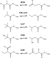
FIGURE 1.
FIGURE 1.
Reactions catalyzed by MCM, ICM, IcmF, HCM, and ECM.
FIGURE 2.
FIGURE 2.
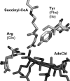
Comparison of active site residues in related AdoCbl-dependent mutases. The MCM structure from Propionibacterium shermanii (Protein Data Bank code 4REQ) was used as a template to show that Tyr and Arg in MCM/ECM correspond to Phe and Gln in ICM/IcmF. In HCM, this pair of residues is Ile and Gln.
FIGURE 3.
FIGURE 3.
Multiple sequence alignment of substrate-binding domains of different AdoCbl-dependent mutases. IcmF from G. kaustophilus (YP_149244), ICM from S. cinnamonensis (AAC08713), MCM from Methylobacterium extorquens (YP_001642233), MeaA from M. extorquens (YP_002961419), ECM from Rhodobacter sphaeroides (YP_354045), and HCM from M. petroleiphilum (YP_001023546) are shown. Four residues recognized to be important for substrate binding are highlighted in gray and indicated with asterisks. Two residues, Phe and Gln, found in ICM and IcmF are substituted by Tyr and Arg in MCM and ECM or Ile and Gln in HCM. ECM is different from other acyl-CoA mutases by the substitution of His and Asp to Gly and Pro, respectively. ECM was previously known as MeaA in M. extorquens. All accession numbers are from NCBI Proteins database.
FIGURE 4.
FIGURE 4.
Generalized scheme for 1,2 rearrangement reaction catalyzed by AdoCbl-dependent mutases. The binding of substrate to the enzyme induces homolytic cleavage of the cobalt-carbon bond of AdoCbl, resulting in formation of a pair of radicals, the 5′-deoxyadenosyl radical and cob(II)alamin. The 5′-deoxyadenosyl radical abstracts a hydrogen atom from the substrate to form 5′-deoxyadenosine and a substrate radical, which subsequently undergoes isomerization to the product radical. The latter reabstracts a hydrogen atom from the methyl group of 5′-deoxyadenosine to form the product and regenerates the 5′-deoxyadenosyl radical. Finally, cob(II)alamin and the 5′-deoxyadenosyl radicals recombine to give AdoCbl.
FIGURE 5.
FIGURE 5.
Organization of genes in mmgABC operon harboring icmF gene. Top, A. flavithermus; middle, B. megaterium; bottom, B. brevis. The following genes are found in the same operon with icmF: tetR (TetR/Acr-like family transcriptional regulator), acdA (acyl-CoA dehydrogenase), mmgA (acetyl-CoA transferase), mmgB (3-OH-butyryl-CoA dehydrogenase), mmgC (acetyl-CoA dehydrogenase), and fadF (medium-/long-chain fatty acyl-CoA dehydrogenase). rpoE gene, which encodes the σ subunit of RNA polymerase, is found just downstream of icmF. The numbers below the genes indicate the distance in nucleotides between two adjacent genes. Negative numbers indicate overlapping genes. For a complete list of bacteria showing similar operonic organization, see the text.
FIGURE 6.
FIGURE 6.
Pivalyl-CoA mutase activity of IcmF. A representative GC chromatogram showing separation of isovaleric and pivalic acids following enzymatic conversion of isovaleryl-CoA to pivalyl-CoA is shown. The reaction mixture contained in Buffer A 15 mm MgCl2, 2000 μg of Gk IcmF, 100 μm AdoCbl, 5 mm GTP, and 1.56 mm isovaleryl-CoA. At different time points, aliquots were removed, the esters were hydrolyzed, and the corresponding acids were separated by GC as described under “Experimental Procedures.” The traces represent 1 min (black line), 5 min (light gray line), 25 min (dark gray line), and pivalic acid standard (dashed line). Valeric acid was used as an internal standard.
FIGURE 7.
FIGURE 7.
Spectral changes in Gk holo-IcmF in presence of isobutyryl-CoA and isovaleryl-CoA. A, spectra of holo-IcmF (40 μm) in buffer A at 24 °C (solid line) and after addition of 4.1 mm isobutyryl-CoA, which resulted in formation of cob(II)alamin (dotted line). B, spectra of holo-IcmF (31 μm) in Buffer A at 24 °C (solid line) and after addition of 1.5 mm isovaleryl-CoA (dotted line).
FIGURE 8.
FIGURE 8.
Inactivation of Gk IcmF during turnover with isobutyryl-CoA. Rapid oxidation of cob(II)alamin to OH2Cbl was observed during reaction of holo-IcmF (with 1 eq; 41 μm AdoCbl bound) with 1.4 mm isobutyryl-CoA in Buffer A with 5 mm MgCl2 at 24 °C in the dark. The spectra were recorded between 0 and 60 min. Inset, the time-dependent increase at 350 nm was fitted to a single exponential function in the absence of nucleotides (filled circles) (kobs = 0.11 ± 0.01 min−1) or in the presence of 5 mm GTP (open circles) (kobs = 0.063 ± 0.002 min−1) or 5 mm ATP (triangles) (kobs = 0.10 ± 0.01 min−1).
FIGURE 9.
FIGURE 9.
Effect of nucleotides on time course of reactions catalyzed by IcmF. A, time course of the isobutyryl-CoA mutase reaction catalyzed by Gk IcmF at 37 °C. The assay mixture in Buffer A with 10 mm MgCl2 contained 40 μg of holo-IcmF, 100 μm AdoCbl, 1.5 mm isobutyryl-CoA, and either no nucleotides (black circles), 6 mm ATP (triangles), or 3 mm GTP (open circles). B, time course of the isovaleryl-CoA mutase reaction catalyzed by Gk IcmF. The reaction mixture in Buffer A with 15 mm MgCl2 contained 2500 μg of IcmF, 100 μm AdoCbl, and 1.5 mm isovaleryl-CoA with (white circles) or without 5 mm GTP (black circles) at 37 °C. Aliquots of the reactions were removed at different time points and analyzed by GC as described under “Experimental Procedures.” Data represent the average of three independent experiments. The data represent the mean ± S.D. of three independent experiments.
FIGURE 10.
FIGURE 10.
Inactivation of Gk IcmF under anaerobic conditions. A, spectral changes upon incubation of 33 μm holo-IcmF (containing 1 eq of bound AdoCbl) with 4.8 mm n-butyryl-CoA in Buffer A with 5 mm MgCl2 under anaerobic conditions at 24 °C in the dark. Spectra were recorded at time 0 (black), immediately after addition of substrate (light gray), and 60 min after addition of substrate (dark gray). Formation of cob(II)alamin (480 nm peak) without further conversion to OH2Cbl was observed. B, comparison of time courses for the reaction catalyzed by Gk IcmF at 37 °C under aerobic (circles) and anaerobic (triangles) conditions in the presence of GTP. The aerobic and anaerobic assay mixtures in Buffer A with 15 mm MgCl2 contained 40 μg of IcmF, 2 mm n-butyryl-CoA, and either no nucleotides (solid symbols) or 4.3 mm GTP (open symbols). The data represent the mean ± S.D. of three independent experiments.
FIGURE 11.
FIGURE 11.
Formation of OH2Cbl and 5′-deoxyadenosine during enzyme-monitored turnover. Holo-IcmF (64 μm IcmF active site concentration containing 2 eq of AdoCbl) was mixed with 1.5 mm isobutyryl-CoA in Buffer A at 37 °C. All manipulations with the samples and HPLC were performed in the dark. The decay of AdoCbl (open triangles) and the appearance of OH2Cbl (solid triangles) and of 5′-deoxyadenosine (open circles) were monitored over 50 min. As a control for AdoCbl stability during sample handling, the analysis was repeated without addition of isobutyryl-CoA (open squares). The data were fitted by a single exponential function for the disappearance of AdoCbl and appearance of 5′-deoxyadenosine and OH2Cbl.
FIGURE 12.
FIGURE 12.
Multiple sequence alignment of IcmFs and MeaB from M. extorquens showing base specificity loop NKX(D/E). Accession numbers are as follows: G. kaustophilus, YP_149244; C. metallidurans CH34, YP_582365; R. eutropha H16, YP_724799; Frankia alni, YP_716016; Nocardia farcinica, YP_117245; B. coagulans, ZP_01696637; Thauera sp., ZP_02841697; Rubrivivax gelatinosus, ZP_00242991; and MeaB, AAL86727. Asp (D) substitution by Glu (E) is indicated by an asterisk. All accession numbers are from the NCBI protein database.

Similar articles

Cited by

References

    1. Banerjee R. (2003) Radical carbon skeleton rearrangements: catalysis by coenzyme B12-dependent mutases. Chem. Rev. 103, 2083–2094 - PubMed
    1. Ratnatilleke A., Vrijbloed J. W., Robinson J. A. (1999) Cloning and sequencing of the coenzyme B12-binding domain of isobutyryl-CoA mutase from Streptomyces cinnamonensis, reconstitution of mutase activity, and characterization of the recombinant enzyme produced in Escherichia coli. J. Biol. Chem. 274, 31679–31685 - PubMed
    1. Cracan V., Padovani D., Banerjee R. (2010) IcmF is a fusion between the radical B12 enzyme isobutyryl-CoA mutase and its G-protein chaperone. J. Biol. Chem. 285, 655–666 - PMC - PubMed
    1. Vrijbloed J. W., Zerbe-Burkhardt K., Ratnatilleke A., Grubelnik-Leiser A., Robinson J. A. (1999) Insertional inactivation of methylmalonyl coenzyme A (CoA) mutase and isobutyryl-CoA mutase genes in Streptomyces cinnamonensis: influence on polyketide antibiotic biosynthesis. J. Bacteriol. 181, 5600–5605 - PMC - PubMed
    1. Rohwerder T., Breuer U., Benndorf D., Lechner U., Müller R. H. (2006) The alkyl tert-butyl ether intermediate 2-hydroxyisobutyrate is degraded via a novel cobalamin-dependent mutase pathway. Appl. Environ. Microbiol. 72, 4128–4135 - PMC - PubMed

Publication types

MeSH terms

LinkOut - more resources