. 2012 Jan 27;287(5):3029-41.
doi: 10.1074/jbc.M111.293985. Epub 2011 Dec 13.

Endogenous vascular endothelial growth factor-A (VEGF-A) maintains endothelial cell homeostasis by regulating VEGF receptor-2 transcription

Affiliations

Endogenous vascular endothelial growth factor-A (VEGF-A) maintains endothelial cell homeostasis by regulating VEGF receptor-2 transcription

Guangqi E et al. J Biol Chem. .

Abstract

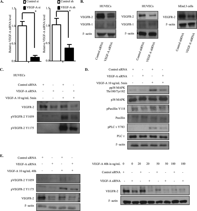
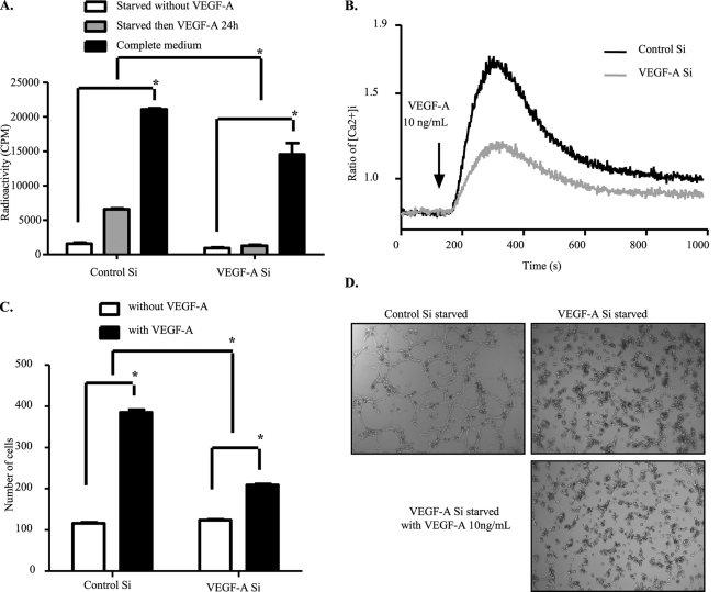
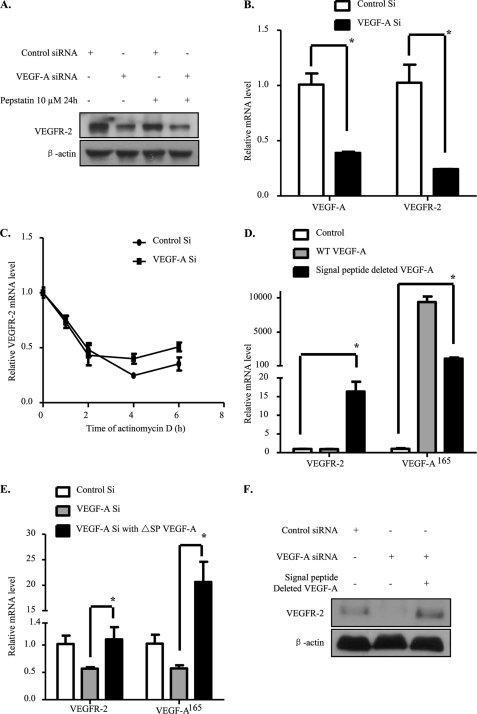
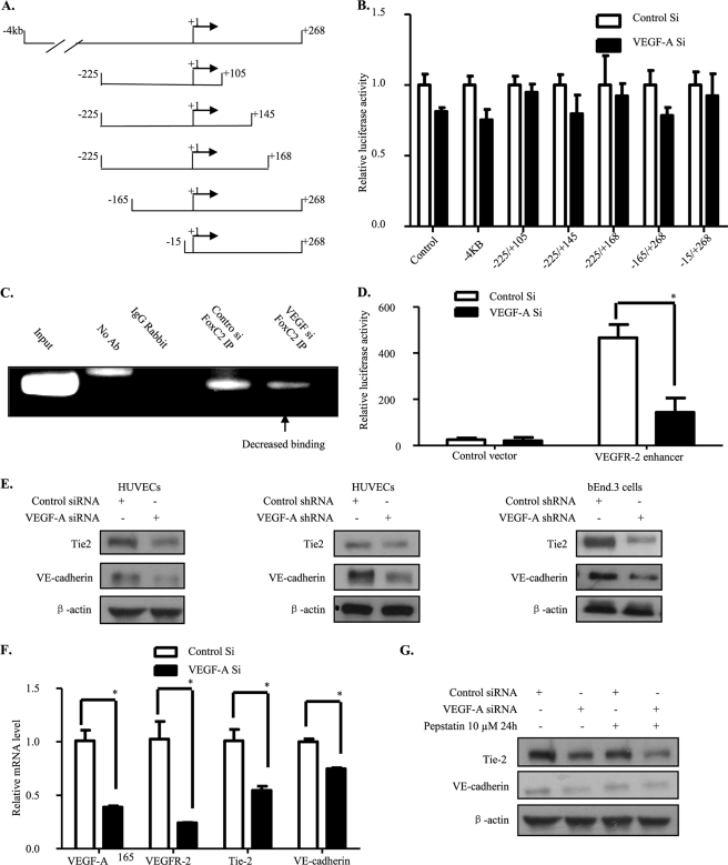
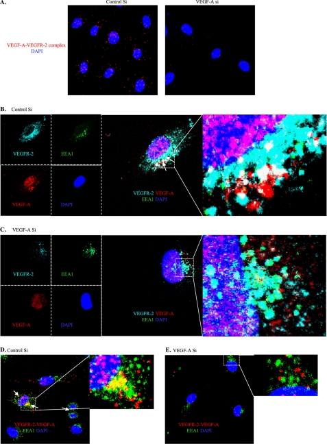
Vascular endothelial growth factor A (VEGF-A) is one of the most important factors controlling angiogenesis. Although the functions of exogenous VEGF-A have been widely studied, the roles of endogenous VEGF-A remain unclear. Here we focused on the mechanistic functions of endogenous VEGF-A in endothelial cells. We found that it is complexed with VEGF receptor 2 (VEGFR-2) and maintains a basal expression level for VEGFR-2 and its downstream signaling activation. Endogenous VEGF-A also controls expression of key endothelial specific genes including VEGFR-2, Tie-2, and vascular endothelial cadherin. Of importance, endogenous VEGF-A differs from exogenous VEGF-A by regulating VEGFR-2 transcription through mediation of FoxC2 binding to the FOX:ETS motif, and the complex formed by endogenous VEGF-A with VEGFR-2 is localized within the EEA1 (early endosome antigen 1) endosomal compartment. Taken together, our results emphasize the importance of endogenous VEGF-A in endothelial cells by regulating key vascular proteins and maintaining the endothelial homeostasis.


Figures

FIGURE 1.
FIGURE 1.
Knockdown of endogenous VEGF-A down-regulates VEGFR-2 expression and its downstream signaling activation. A and B, HUVECs were treated with control or VEGF-A siRNA. Forty-eight hours after transfection, cells were lysed and analyzed by Western blot. For shRNA, HUVECs or bEnd.3 cells were infected by lentivirus for 24 h, selected by puromycin for 48 h, and lysed. For mRNA analysis, mRNA were extracted and then reverse-transcribed into cDNA. The latter was used for real-time PCR. C and D, siRNA-transfected HUVECs were starved overnight and stressed by VEGF-A165 at 10 ng/ml for 10 min and then quenched and lysed. E, HUVECs were treated by control or VEGF-A siRNA. Eight hours later, VEGF-A165 was added into the culture medium, and the cells incubated for 40 h. The cells were then lysed and analyzed by Western blot.
FIGURE 2.
FIGURE 2.
Endothelial functions displayed by VEGFR-2 are impaired upon depletion of VEGF-A. A, shown is the reduced proliferation capacity of endogenous VEGF-A knockdown cells. HUVECs were transfected with control or VEGF-A siRNA for 48 h, and cell proliferation was then measured by [3H]thymidine incorporation assay. B, reduction in calcium release upon VEGF-A stimulation is shown. HUVECs were transfected with control or VEGF-A siRNA. Forty-eight hours later cells were digested and loaded with Fura-2. Intracellular calcium release upon VEGF-A stress was followed for 1000 s. C, cell migration capacity was affected by endogenous VEGF-A silencing. siRNA-treated cells were starved overnight. Forty-eight hours after transfection, cells were seeded into the upper chambers of Transwells and allowed to migrate into the bottom chambers containing VEGF-A165 (10 ng/ml) as the chemoattractant. Four hours later, cells were fixed and photographed for counting. D, tube formation was inhibited by depletion of endogenous VEGF-A. siRNA-transfected HUVECs were starved overnight, then seeded onto solidified Matrigel. VEGF-A165 (10 ng/ml) was then added into the culture medium, and tube formation was followed for 8 h. The data represented here are the average of three independent experiments. All bars represent means ± S.D. of three experiments. *, p < 0.05.
FIGURE 3.
FIGURE 3.
VEGFR-2 decrease is due to transcriptional down-regulation. A, VEGFR-2 down-regulation was not due to lysosomal degradation. siRNA-transfected HUVECs were treated with pepstatin at 10 μm for 24 h. Forty-eight hours after transfection cells were lysed and analyzed by Western blot. B, the VEGFR-2 mRNA level was decreased. Forty-eight hours after siRNA transfection, HUVECs were harvested for mRNA extraction. cDNA was obtained by reverse transcription, and real-time PCR was performed. C, mRNA stability was unaffected. Forty-eight hours after siRNA transfection, cells were treated with 7.5 μg/ml actinomycin D at different time points, the mRNA was harvested, and the procedure was repeated. The data represented here are the average of three independent results. D, a non-secretive form of VEGF-A up-regulates VEGFR-2 mRNA level. HUVECs were infected by retroviral overexpression system, and 48 h later mRNAs were extracted. Real-time PCR were realized with reverse-transcribed cDNA. E and F, HUVECs were first transfected by siRNA and 6 h later a retrovirus overexpressing non-secretive form of VEGF-A was added. 72 h after siRNA treatment, mRNAs were extracted for real-time PCR or cells were lysed for Western blot. All bars represent the means ± S.D. of three experiments. *, p < 0.05.
FIGURE 4.
FIGURE 4.
FoxC2 binding to the FOX:ETS motif is decreased. A, shown is a representation of VEGFR-2 promoter deletion mutants used in our studies. B, VEGFR-2 promoter activation was unaffected by deletions. Different VEGFR-2 promoter deletion-mutant luciferase constructs with Renilla luciferase vector and control or VEGF-A siRNA were co-transfected into HUVECs. Forty-eight hours later, firefly and Renilla luciferase activities were measured. C, binding of FoxC2 to the FOX:ETS motif was decreased by endogenous VEGF-A knockdown. Forty-eight hours after siRNA transfection, HUVECs were subjected to chromatin immunoprecipitation by anti-FoxC2 antibody. Immunoprecipitated DNA fragments were used to perform PCR. D, pGL3-promoter empty vector or containing VEGFR-2 enhancer constructs with Renilla luciferase vector and control or VEGF-A siRNA were co-transfected into HUVECs. Forty-eight hours later, firefly and Renilla luciferase activities were measured. E–G, VE-cadherin and Tie-2 levels were also down-regulated. The same procedure as for VEGFR-2 in Fig. 3 was followed. All bars represent means ± S.D. of three experiments. *, p < 0.05.
FIGURE 5.
FIGURE 5.
Endogenous VEGF-A binds to VEGFR-2 and is partially localized within the EEA1 endosomal compartment. A, endogenous VEGF-A binds to VEGFR-2. In situ PLA was performed for VEGFR-2 and VEGF-A on siRNA-transfected HUVECs. B–E, shown is co-distribution of the VEGF-A-VEGFR-2 complex with EEA1. White arrows indicate colocalization zones. B and C, immunofluorescence was performed with HUVECs for VEGF-A (red), VEGFR-2 (far red), and EEA1 (green). D and E, in situ PLA of VEGFR-2-VEGF-A was co-stained with EEA1-FITC.

References

    1. Senger D. R., Galli S. J., Dvorak A. M., Perruzzi C. A., Harvey V. S., Dvorak H. F. (1983) Tumor cells secrete a vascular permeability factor that promotes accumulation of ascites fluid. Science 219, 983–985 - PubMed
    1. Dvorak H. F., Nagy J. A., Feng D., Brown L. F., Dvorak A. M. (1999) Vascular permeability factor/vascular endothelial growth factor and the significance of microvascular hyperpermeability in angiogenesis. Curr. Top. Microbiol. Immunol. 237, 97–132 - PubMed
    1. Nagy J. A., Vasile E., Feng D., Sundberg C., Brown L. F., Manseau E. J., Dvorak A. M., Dvorak H. F. (2002) VEGF-A induces angiogenesis, arteriogenesis, lymphangiogenesis, and vascular malformations. Cold Spring Harb. Symp. Quant. Biol. 67, 227–237 - PubMed
    1. Pettersson A., Nagy J. A., Brown L. F., Sundberg C., Morgan E., Jungles S., Carter R., Krieger J. E., Manseau E. J., Harvey V. S., Eckelhoefer I. A., Feng D., Dvorak A. M., Mulligan R. C., Dvorak H. F. (2000) Heterogeneity of the angiogenic response induced in different normal adult tissues by vascular permeability factor/vascular endothelial growth factor. Lab. Invest. 80, 99–115 - PubMed
    1. Benjamin L. E., Keshet E. (1997) Conditional switching of vascular endothelial growth factor (VEGF) expression in tumors. Induction of endothelial cell shedding and regression of hemangioblastoma-like vessels by VEGF withdrawal. Proc. Natl. Acad. Sci. U.S.A. 94, 8761–8766 - PMC - PubMed

Publication types

MeSH terms

Substances