. 2012 Feb 3;287(6):4275-87.
doi: 10.1074/jbc.M111.280016. Epub 2011 Dec 13.

Ex vivo and in vivo effects of isofagomine on acid β-glucosidase variants and substrate levels in Gaucher disease

Affiliations

Ex vivo and in vivo effects of isofagomine on acid β-glucosidase variants and substrate levels in Gaucher disease

Ying Sun et al. J Biol Chem. .

Abstract

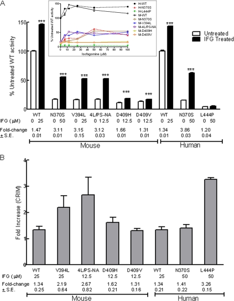
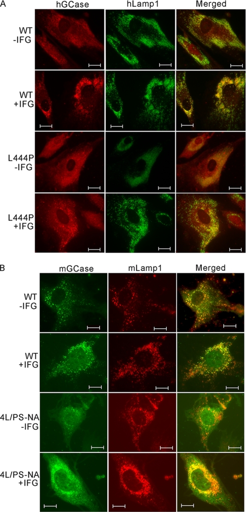
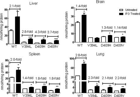
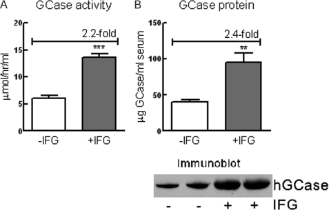
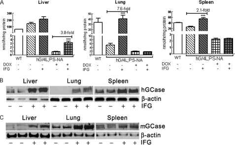
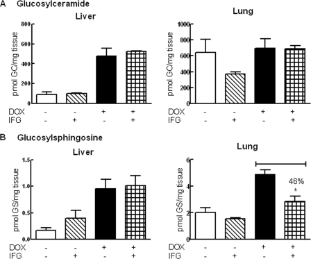
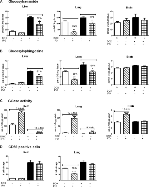
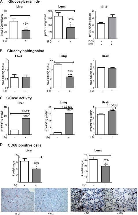
Isofagomine (IFG) is an acid β-glucosidase (GCase) active site inhibitor that acts as a pharmacological chaperone. The effect of IFG on GCase function was investigated in GCase mutant fibroblasts and mouse models. IFG inhibits GCase with K(i) ∼30 nM for wild-type and mutant enzymes (N370S and V394L). Fibroblasts treated with IFG at μM concentrations showed enhancement of WT and mutant GCase activities and protein levels. Administration of IFG (30 mg/kg/day) to the mice homozygous for GCase mutations (V394L, D409H, or D409V) led to increased GCase activity in visceral tissues and brain extracts. IFG effects on GCase stability and substrate levels were evaluated in a mouse model (hG/4L/PS-NA) that has doxycycline-controlled human WT GCase (hGCase) expression driven by a liver-specific promoter and is also homozygous for the IFG-responsive V394L GCase. Both human and mouse GCase activity and protein levels were increased in IFG-treated mice. The liver-secreted hGCase in serum was stabilized, and its effect on the lung and spleen involvement was enhanced by IFG treatment. In 8-week IFG-treated mice, the accumulated glucosylceramide and glucosylsphingosine were reduced by 75 and 33%, respectively. Decreases of storage cells were correlated with >50% reductions in substrate levels. These results indicate that IFG stabilizes GCase in tissues and serum and can reduce visceral substrates in vivo.


Figures

FIGURE 1.
FIGURE 1.
IFG effects on fibroblast GCase activity (A) and protein (B) levels. A, mouse (M) (WT, N370S, V394L, 4L/PS-NA, D409H, and D409V) and human (H) (WT, N370S, and L444P) fibroblast cells were incubated with IFG. GCase activity at the indicated concentration to achieve peak effects (see inset) is presented as the percentage of untreated WT activity. All treated mouse and human WT and mutant cells except L444P showed significant increases of activity. The activities in treated mouse V394L and 4L/PS-NA increased to ∼50% of WT levels. GCase activity in IFG-treated human N370S cells was enhanced to 60% of WT levels. -Fold changes relative to the untreated cells are indicated. The experiments were done in triplicate. Data were analyzed by Student's t test. ***, p < 0.001. Inset, IFG concentration-response curves for human and mouse WT and mutant GCases in fibroblasts. The cells were treated with IFG at the indicated concentrations for 5 days. GCase activity changes after IFG treatment are presented as a percentage of untreated WT activity for mouse or human, respectively. B, quantitation of GCase protein in each variant was determined immunologically with mouse- or human-specific antibodies. All IFG-treated cells showed increases in GCase protein levels. These are presented as -fold change (indicated below the columns) relative to the untreated cells. The results and error bars represent the mean ± S.E. (n = 3).
FIGURE 2.
FIGURE 2.
Cellular localization of GCases in IFG-treated fibroblasts. Colocalization of hGCase (A) (red) with lysosomal marker human Lamp 1 (green) and mouse GCase (mGCase) (B) (green) with mouse Lamp1 (red) was conducted by immunofluorescence staining. The fibroblast cells were treated with IFG (50 μm). Both untreated (−IFG) and treated (+IFG) human and mouse WT GCases colocalized with Lamp1 in the lysosome. The untreated human L444P GCase was nearly undetectable. In contrast, the treated L444P cells had increased GCase and trafficking to the lysosome. In IFG-treated mouse 4L/PS-NA cells, the GCase signal was enhanced and localized in the lysosome. Scale bars, 10 μm.
FIGURE 3.
FIGURE 3.
IFG treatment enhanced GCase activity in mouse tissues. The mice were administered IFG (30 mg/kg/day) in drinking water. The treatments for WT and V394L mice were started at postnatal day 10 and continued for 5 weeks; for D409H mice, the treatment started at 21 days and continued for 4 weeks; and for D409V mice, the treatment started at 23 days and continued for 8 weeks. GCase activities in each tissue were significantly increased in IFG-treated WT and mutant mice tissues except V394L brain. Data were analyzed by Student's t test. *, p < 0.05; **, p < 0.01; ***, p < 0.001. The results and error bars represent the mean ± S.E. (n = 3 mice).
FIGURE 4.
FIGURE 4.
IFG stabilized GCase protein in serum. The hGCase is expressed in and secreted from hG/4L/PS-NA mouse livers. A, in IFG-treated mice (30 mg/kg/day for 4 weeks) hGCase activities secreted into the serum were significantly increased compared with untreated mice. B, immunoblot of GCase in serum determined by anti-hGCase antibody and quantitated relative to known amounts of imiglucerase as standard. Human GCase protein in the serum from IFG-treated mice was significantly increased compared with the untreated mice. Data were analyzed by Student's t test (duplicate assays). **, p < 0.01; ***, p < 0.001. The results and error bars represent the mean ± S.E. (n = 3 mice).
FIGURE 5.
FIGURE 5.
GCase activity and protein in IFG-treated hG/4L/PS-NA mice. Human GCase was expressed in the liver of hG/4L/PS-NA mice when fed DOX-free food. In the mice on DOX food, hGCase expression was turned off; residual activity was from mouse V394L mutant GCase. A, IFG treatment for 4 weeks in drinking water enhanced both hGCase and mouse V394L mutant GCase activities in the liver and hGCase activity in the lung and spleen (gray bars). B, immunoblot using anti-hGCase antibody demonstrated that IFG treatment for 4 weeks increased hGCase protein levels in the liver, lung, and spleen of hG/4L/PS-NA mice. No protein was detected in untreated lung. C, immunoblot of mouse GCase detected by anti-mouse GCase antibody showed increases of mouse GCase (mGCase) protein in treated liver, lung, and spleen. β-Actin was the loading control. Data were analyzed by Student's t test. ***, p < 0.001. The results and error bars represent the mean ± S.E. (n = 3 mice).
FIGURE 6.
FIGURE 6.
Effects of IFG treatment for 4 weeks in hG/4L/PS-NA mice. The mice were treated with IFG for 4 weeks with DOX-free or DOX food. A, no significant changes of GC levels were observed in IFG-treated liver and lung. B, in liver of DOX-free IFG-treated mice, GS was increased. In lungs of DOX and IFG-treated mice, GS was decreased. The liver GS level was not altered in these same mice. GC and GS levels were quantitated by LC/MS and normalized by mg of wet tissues. Data were analyzed by Student's t test. *, p < 0.05. GCase activities for these samples are presented in Fig. 5. The results and error bars represent the mean ± S.E. (n = 3 mice).
FIGURE 7.
FIGURE 7.
Effects of IFG treatment for 8 weeks in hG/4L/PS-NA mice. IFG was administered to the mice by drinking water for 8 weeks with the mice either on DOX or DOX-free food. A, the liver GC level was low in mice on DOX-free food and not significantly changed by IFG treatment. The liver GC in the mice fed with DOX food was reduced by 38% with IFG treatment. In comparison, lung GC levels were significantly reduced in IFG-treated mice on either DOX or DOX-free food. Brain GC levels were not significantly changed by IFG treatment. B, the liver GS level was very low in the mice on DOX-free food and not significantly altered by IFG, whereas in mice fed DOX food, GS was reduced by 39% with IFG treatment. Lung GS levels were significantly reduced in IFG-treated mice on DOX or DOX-free food. Brain GS levels were not significantly changed by IFG treatment. C, GCase activity. IFG treatment for 8 weeks in drinking water enhanced both hGCase (hatched bars) and mouse V394L mutant GCase (checked bars) activities in the liver and lung. IFG treatment enhanced hGCase activity (hatched bars) by 1.6-fold in brain but did not alter mouse V394L GCase activity (checked bars). D, the CD68-positive macrophages were counted in liver and lung sections of 8-week IFG-treated mice. Decreased cell numbers were observed in IFG-treated lungs from the mice on DOX-free food. The CD68-positive cells were counted from 10 images (305 × 228 μm for each image) per mouse. GC and GS levels were quantitated by LC/MS and normalized by mg of wet tissues. Data were analyzed by Student's t test. *, p < 0.05; **, p < 0.01; ***, p < 0.001, ****, p < 0.0001. The results and error bars represent the mean ± S.E. (n = 3 mice).
FIGURE 8.
FIGURE 8.
Reduced substrates and storage cells in hG/4L/PS-NA mice treated for 4 weeks with IFG during hGCase synthesis in the liver. hG/4L/PS-NA mice were placed on DOX food from 3 to 7 weeks of age to turn off hGCase expression. The mice were then withdrawn from DOX to permit hGCase synthesis and fed with IFG-containing drinking water from 7 to 11 weeks of age. A, GC levels in IFG-treated liver and lung were reduced. Brain GC levels were not significantly changed by IFG treatment. B, GS levels were decreased in IFG-treated mouse lungs. C, GCase activities were increased in IFG-treated liver, lung, and brain. D, CD68-positive cell numbers in IFG-treated mice were decreased in both liver (upper panel) and lung (lower panel). Images of liver and lung were stained with anti-CD68 antibody on macrophage cells (brown) counterstained with hematoxylin (blue). The cell counts and lipid level determination are as described in Fig. 7. Data were analyzed by Student's t test. *, p < 0.05; **, p < 0.01; ***, p < 0.001, ****, p < 0.0001. The results and error bars represent the mean ± S.E. (n = 3 mice).

References

    1. Grabowski G. A., Petsko G., Kolodny E. H. (2010) in The Online Metabolic and Molecular Bases of Inherited Diseases (Valle D., Beaudet A. L., Vogelstein B., Kinzler K. W., Antonarakis S. E., Ballabio A., Scriver C. R., Sly W. S., Childs B., eds) 9th Ed., Chapter 146, McGraw-Hill, New York
    1. Koprivica V., Stone D. L., Park J. K., Callahan M., Frisch A., Cohen I. J., Tayebi N., Sidransky E. (2000) Analysis and classification of 304 mutant alleles in patients with type 1 and type 3 Gaucher disease. Am. J. Hum. Genet. 66, 1777–1786 - PMC - PubMed
    1. Horowitz M., Wilder S., Horowitz Z., Reiner O., Gelbart T., Beutler E. (1989) The human glucocerebrosidase gene and pseudogene: structure and evolution. Genomics 4, 87–96 - PubMed
    1. Kolodny E. H., Ullman M. D., Mankin H. J., Raghavan S. S., Topol J., Sullivan J. L. (1982) Phenotypic manifestations of Gaucher disease: clinical features in 48 biochemically verified type 1 patients and comment on type 2 patients. Prog. Clin. Biol. Res. 95, 33–65 - PubMed
    1. Volk B. W., Wallace B. J., Adachi M. (1967) Infantile Gaucher's disease: electron microscopic and histochemical studies of a cerebral biopsy. J. Neuropathol. Exp. Neurol. 26, 176–177 - PubMed

MeSH terms