Comparative Study
. 2012 Jan 24;109(4):1210-5.
doi: 10.1073/pnas.1118834109. Epub 2011 Dec 13.

Natural killer cell activation enhances immune pathology and promotes chronic infection by limiting CD8+ T-cell immunity

Affiliations
Comparative Study

Natural killer cell activation enhances immune pathology and promotes chronic infection by limiting CD8+ T-cell immunity

Philipp A Lang et al. Proc Natl Acad Sci U S A. .

Abstract

Infections with HIV, hepatitis B virus, and hepatitis C virus can turn into chronic infections, which currently affect more than 500 million patients worldwide. It is generally thought that virus-mediated T-cell exhaustion limits T-cell function, thus promoting chronic disease. Here we demonstrate that natural killer (NK) cells have a negative impact on the development of T-cell immunity by using the murine lymphocytic choriomeningitis virus. NK cell-deficient (Nfil3(-/-), E4BP4(-/-)) mice exhibited a higher virus-specific T-cell response. In addition, NK cell depletion caused enhanced T-cell immunity in WT mice, which led to rapid virus control and prevented chronic infection in lymphocytic choriomeningitis virus clone 13- and reduced viral load in DOCILE-infected animals. Further experiments showed that NKG2D triggered regulatory NK cell functions, which were mediated by perforin, and limited T-cell responses. Therefore, we identified an important role of regulatory NK cells in limiting T-cell immunity during virus infection.


Conflict of interest statement

The authors declare no conflict of interest.

Figures

Fig. 1.
Fig. 1.
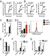
NK cells are activated after LCMV infection. (A) Splenocytes from naïve (Left), LCMV-Armstrong–infected (Center), and LCMV clone 13-infected (Right) mice were incubated with YAC-1 cells at the indicated effector/target ratios. 51Cr release at different effector/target ratios is shown (n = 3, mean ± SEM of duplicates). (B and C) NK1.1+CD3e splenocytes were analyzed by flow cytometry for surface expression of NKG2D (B) and 2B4 (C) at 2 d after infection with 2 × 10⁶ pfu of LCMV WE (n = 6; Student's t test). NKG2D expression (mean fluorescence intensity) (B) and 2B4 negative (%) (C) NK1.1+CD3 cells are shown (n = 6; Student's t test).
Fig. 2.
Fig. 2.
NK cells limit CD8+ T-cell response in vivo and in vitro. WT and NK cell-depleted mice were infected with 2 × 10⁶ pfu of LCMV WE. (A) gp33-specific (Left) and np396-specific (Right) tetramer response in the spleen (% of CD8+ T cells) at the indicated time (days) after infection are shown (P < 0.01 for day 6 and P < 0.001 for day 8 gp33 tetramers; P < 0.001 for day 6 and 8 for np396 tetramers; two-way ANOVA, n = 6 of two independent experiments). (B) IFN-γ+CD8+ of all CD8+ splenocytes are shown after restimulation with gp33 (Left) or np396 (Right) peptide (P < 0.001 for days 6 and 8 postinfection, two-way ANOVA, n = 6). (C) Control or NK cell-depleted mice were infected with 200 pfu of LCMV WE. At 8 d postinfection, splenocytes were restimulated with the LCMV-specific epitope gp33, and IFN-γ production was determined (n = 7–8; two-way ANOVA). (D) A total of 2 × 10⁶ negatively sorted, CD45.1+ P14 T cells were transferred at 2 d postinfection with 200 pfu of LCMV WE into control and NK cell-depleted mice. CD45.1+CD8+ population was analyzed (Left; n = 6; P < 0.05 for aNK1.1 from naïve and control group using one-way ANOVA), and NKG2D–hIgG expression was measured (one representative of n = 3–6 is shown). (E) Negatively sorted T cells were activated with anti-CD3/CD28 antibodies and coincubated with purified NK cells from naïve or LCMV-infected mice (day 2, 2 × 10⁶ pfu) or naïve NK cells stimulated with IFN-α4. CD8+ T-cell number was compared after 72 h (n = 3–4; *P < 0.05, one-way ANOVA). (F) Nfil3−/− and nfil3+/− mice were infected with 2 × 10⁵ pfu of LCMV WE. At 6 d after infection, splenocytes were restimulated in vitro with the virus-specific peptides gp33 and np396, and IFN-γ production was determined by intracellular cytokine staining and FCM analysis (n = 5–6 of two independent experiments; P < 0.01 for gp33 and np396, two-way ANOVA). (G) WT or NK cell-depleted mice were infected with 2 × 10⁶ pfu of LCMV clone 13. Splenocytes from control or NK cell-depleted mice were restimulated ex vivo with the virus-derived peptides gp33 and np396 at 10 d after infection. Intracellular production of IFN-γ by CD8+ T cells was measured by FCM analysis (n = 6 of two independent experiments; P < 0.001 for gp33 and np396, two-way ANOVA).
Fig. 3.
Fig. 3.
NKG2D triggers NKreg functions. (A) Mice were treated with NKG2D-blocking antibody or isotype control (day −1, day 2 postinfection) and infected with 200 pfu of LCMV WE (day 0). On day 2, 2 × 10⁶ negatively sorted, CFSE-labeled, and congenically marked P14 T cells were transferred into animals of both groups. On day 4 after infection, CFSE expression and expansion of CD45.1+CD8+ cells were determined (n = 3–4; Student's t test). (B and C) Mice were treated with NKG2D-blocking antibody or an isotype control (day −1, 12 h postinfection) and infected with 2 × 10⁶ pfu of LCMV WE. gp33-tetramer+ was measured at 8 d after infection (n = 3; Student's t test) (B), and IFN-γ production was determined after restimulation with the virus-specific epitopes gp33 and np396 (C) (n = 3; two-way ANOVA).
Fig. 4.
Fig. 4.
NK cell-mediated perforin production limits T-cell immunity. (A and B) Splenocytes (50,000) from P14+CD45.1+ mice were transferred into perforin−/− and WT mice and infected with 2 × 10⁶ pfu of LCMV WE. (A) At 8 d postinfection, IFN-γ production of CD8+ T cells after restimulation with gp33 was determined in single-cell suspensions of spleen and liver tissue (n = 6; P < 0.01, Student's t test). (B) One group was treated with an NK cell-depleting antibody, and IFN-γ production was monitored (n = 5–6; *P < 0.05 for aNK1.1, perforin−/−, and aNK1.1 perforin−/− vs. control, Newman–Keuls one-way ANOVA). (C) A total of 2 × 10⁶ negatively sorted, CFSE-labeled T cells from P14+CD45.1+ mice were transferred into WT and perforin−/− mice on day 2 postinfection with 200 pfu of LCMV WE. (Left) After 48 h, CFSE expression of the CD45.1+CD8+ population was analyzed. (Right) Relative amount (%) of CD45.1+CD8+ cells in perforin−/− mice compared with WT mice (n = 4; P < 0.01, Student's t test).
Fig. 5.
Fig. 5.
NK cells promote development of chronic infection and immunopathology. (A) WT or NK cell-depleted mice were infected with 2 × 10⁶ pfu of LCMV clone 13. Virus titers were determined from spleen, liver, kidney, and lung tissue at 10 d after infection [n = 8 of two independent experiments; P < 0.001 except liver (P < 0.05) of 10log(Titer), Student's t test]. (B) Control and NK cell-depleted mice were infected with 105 pfu of LCMV DOCILE. Virus titers were measured after 33 d (n = 5, Student's t test). (CE) WT or NK cell-depleted mice were infected with 2 × 10⁶ pfu of LCMV WE. (C) At 6 and 8 d postinfection, virus titers were analyzed in spleen tissue. (n = 6). *P < 0.05, significant difference for 10log(Titer), Student's t test. (D) Liver from control and NK cell-depleted mice were analyzed for LCMV nucleoprotein expression by immunohistology. One representative picture is shown (n = 5). (E) WT mice or NK cell-depleted mice were infected with 2 × 10⁶ pfu of LCMV. Hepatitis induction was assessed by measuring ALT activity in the sera at different time points (n = 2–11; P < 0.05 for day 10 and P < 0.01 for day 12 postinfection, two-way ANOVA). (F) Nfil3−/− and nfil3+/− mice were infected with 2 × 10⁵ pfu of LCMV WE. ALT activity in the serum of nfil3−/− and nfil3+/− mice was determined at 11 d postinfection (n = 7–8; P < 0.01 using Student's t test).

Comment in

References

    1. Vivier E, Tomasello E, Baratin M, Walzer T, Ugolini S. Functions of natural killer cells. Nat Immunol. 2008;9:503–510. - PubMed
    1. Walzer T, Dalod M, Robbins SH, Zitvogel L, Vivier E. Natural-killer cells and dendritic cells: “L'union fait la force”. Blood. 2005;106:2252–2258. - PubMed
    1. Sivori S, et al. CpG and double-stranded RNA trigger human NK cells by Toll-like receptors: Induction of cytokine release and cytotoxicity against tumors and dendritic cells. Proc Natl Acad Sci USA. 2004;101:10116–10121. - PMC - PubMed
    1. Biron CA, Nguyen KB, Pien GC. Innate immune responses to LCMV infections: Natural killer cells and cytokines. Curr Top Microbiol Immunol. 2002;263:7–27. - PubMed
    1. Vivier E, Nunès JA, Vély F. Natural killer cell signaling pathways. Science. 2004;306:1517–1519. - PubMed

Publication types

MeSH terms

Substances