Review
. 2011 Nov;4(11-12):773-87.
doi: 10.1002/jbio.201100062.

A review of in-vivo optical properties of human tissues and its impact on PDT

Affiliations
Review

A review of in-vivo optical properties of human tissues and its impact on PDT

Julia L Sandell et al. J Biophotonics. 2011 Nov.

Abstract

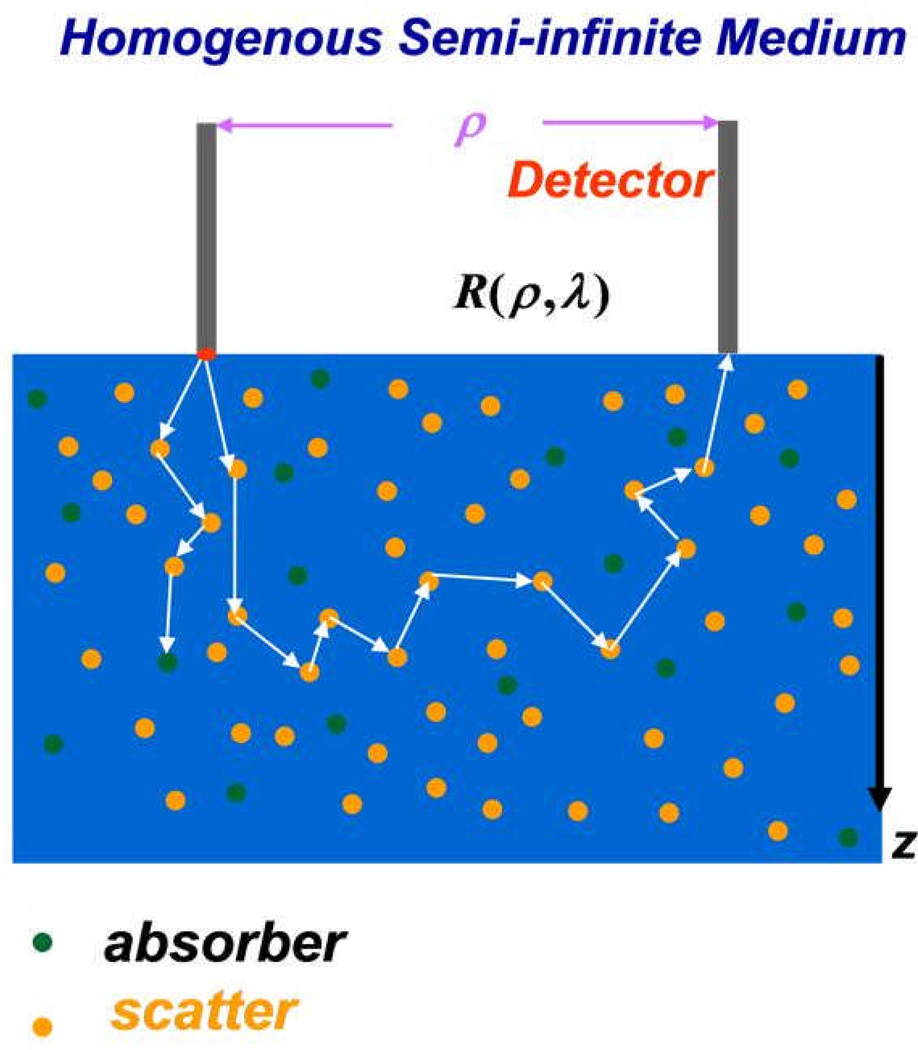
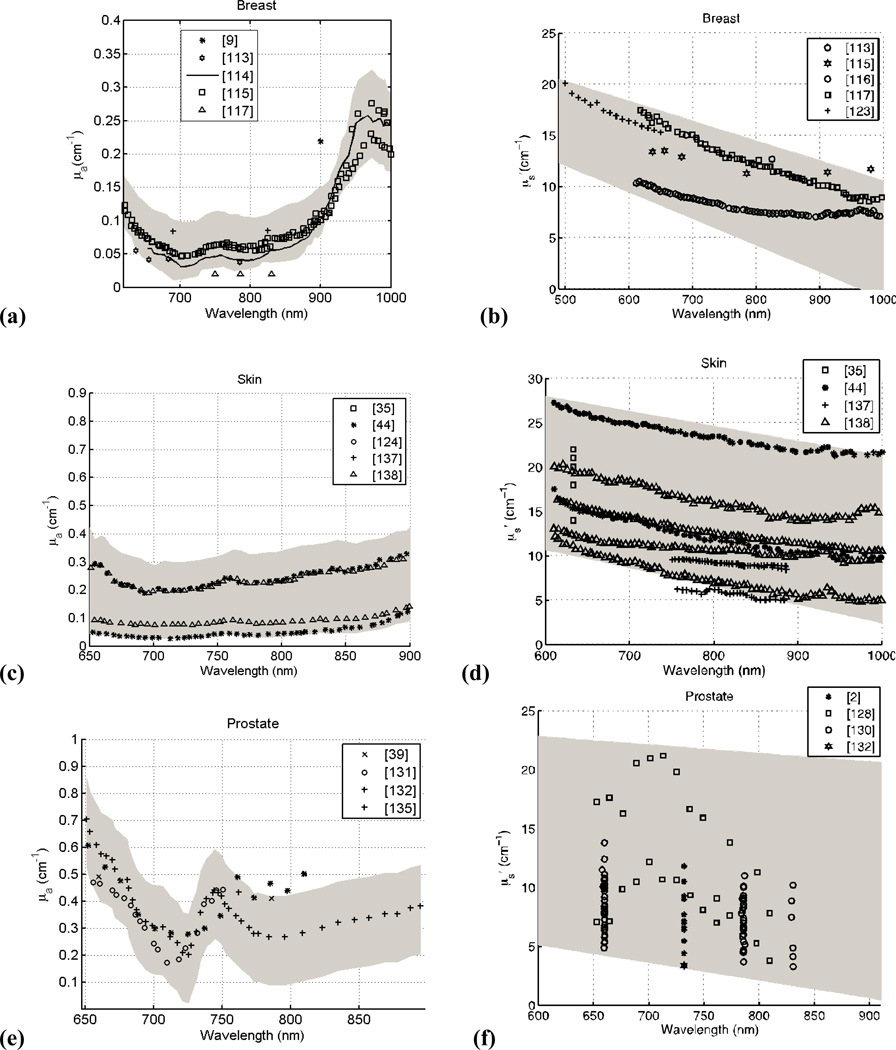
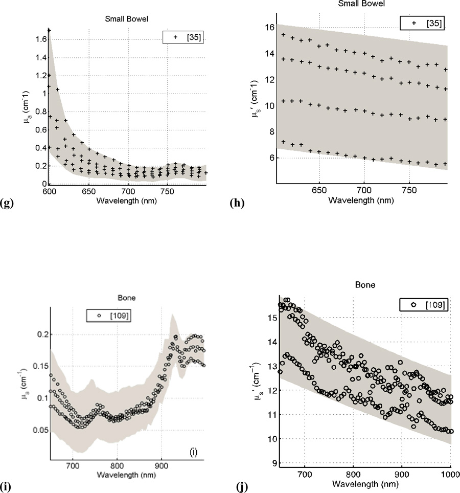
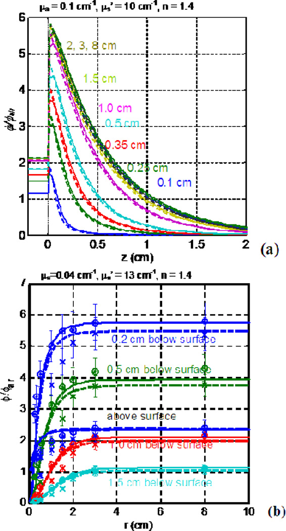
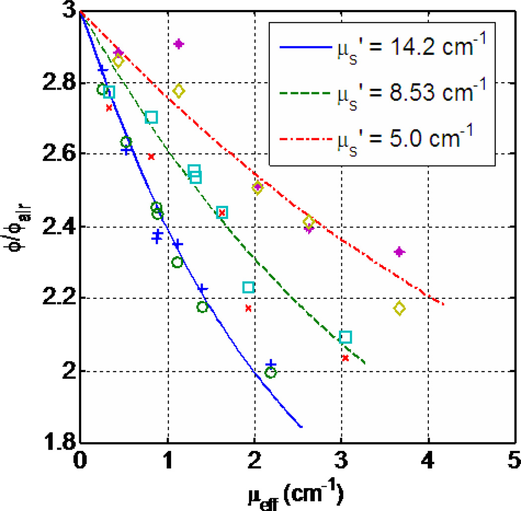
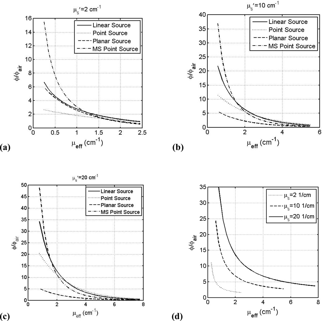
A thorough understanding of optical properties of biological tissues is critical to effective treatment planning for therapies such as photodynamic therapy (PDT). In the last two decades, new technologies, such as broadband diffuse spectroscopy, have been developed to obtain in vivo data in humans that was not possible before. We found that the in vivo optical properties generally vary in the ranges μ(a) = 0.03-1.6 cm⁻¹ and μ'(s) = 1.2-40 cm⁻¹, although the actual range is tissue-type dependent. We have also examined the overall trend of the absorption spectra (for μ(a) and μ'(s)) as a function of wavelength within a 95% confidence interval for various tissues in vivo. The impact of optical properties on light fluence rate is also discussed for various light application geometries including superficial, interstitial, and within a cavity.


Figures

Figure 1
Figure 1
Schematics of broad-band diffuse reflectance spectroscopy (BDRS). Pencil beam light source was incident from the left fiber. The photons are randomly scattered (depicted by orange dots) and absorbed (depicted by green dots) as they travel through tissue. The absorbed photons are trapped in the tissue while the scattered photons continue to either be absorbed or scattered and ultimately detected by the detector fiber located at a distance ρ from the incident light source.
Figure 2
Figure 2
Extrapolation of optical properties from measured transmission data from a point source. The measured data (symbols) are fitted with a best-fit using the optical properties according to Eq. 8 (line); these data are displayed in the lower left-hand panel. Taken from [33].
Figure 3
Figure 3
Molar absorption coefficients of common absorbents in tissue: oxy-hemoglobin, deoxy-hemoglobin, melanin, and water.
Figure 4
Figure 4
Absorption and Scattering coefficients of human tissue in vivo versus wavelength for breast (a, b); skin (c, d); prostate (e, f); small bowel (g, h); and bone (i, j). The grey shaded region is the range of μa and μs’ with a 95% confidence interval based on review of the literature.
Figure 4
Figure 4
Absorption and Scattering coefficients of human tissue in vivo versus wavelength for breast (a, b); skin (c, d); prostate (e, f); small bowel (g, h); and bone (i, j). The grey shaded region is the range of μa and μs’ with a 95% confidence interval based on review of the literature.
Figure 5
Figure 5
MC-calculated ϕ/ϕair as a function of tissue depth (a) and beam radius (b). The ϕ/ϕair was calculated for μa = 0.1 cm−1, μs’ = 10 cm−1, and index of refraction n=1.4 for varying beam radii. Note the exponential decrease with increasing depth. The ϕ/ϕair becomes a constant after the beam radius reaches 2 cm.
Figure 6
Figure 6
Relationship between the diffuse reflectance Rd and optical properties. The symbols are measurements and the lines are theoretical calculations described in the text. Data are taken from Ref. [92].
Figure 7
Figure 7
Fluence rate, normalized to in-air fluence rate ϕair, versus μeff for linear (solid line), point sources (dashed line) inside an infinite medium, planar light sources (dotted line) on a semi-infinite medium below an air-tissue interface, and point source with multiple scattering (MS) (dash-dotted line) inside a spherical cavity of 10 cm radius at 0.5 cm from the light source (for point or linear sources) or 0.5 cm depth in tissue (planar or MS point sources) for μs’: (a) 2.0 cm−1, (b) 10.0 cm−1, (c) 20 cm−1. See text for the definition of ϕair. (d) ϕ/ϕair within a spherical cavity at air-tissue boundary for μs’=2.0 cm−1 (dotted), 10.0 cm−1 (dashed), and 20.0 cm−1 (solid) using Eqs. 10 and 12.

References

    1. Graff R, Dassel ACM, Koelink MH, de Mul F, de Mul FFM, Aarnoudse JG, Zijistra WG. Appl Opt. 1993;32(4):435–447. - PubMed
    1. Zhu TC, Hahn SM, Kapatkin AS, Dimofte A, Rodriguez CE, Vulcan T, Glatstein E, Hsi RA. Photochem Photobiol. 2003;77:81–88. - PubMed
    1. Cheong WF, Prahl SA, Welch AJ. IEEE J Quan Elec. 1990;26(12):2166–2185.
    1. Wilson BC, Jeeves WP, Lowe DM. Photochem Photobiol. 1985;42(2):153–162. - PubMed
    1. Marchesini R, Clemente C, Pignoli E, Brambilla M. J Photochem. Photobiol. 1992;16(2):127–140. - PubMed

Publication types