Review
. 2012 Jan;245(1):189-208.
doi: 10.1111/j.1600-065X.2011.01074.x.

Immune modulation during latent herpesvirus infection

Affiliations
Review

Immune modulation during latent herpesvirus infection

Douglas W White et al. Immunol Rev. 2012 Jan.

Abstract

Nearly all human beings, by the time they reach adolescence, are infected with multiple herpesviruses. At any given time, this family of viruses accounts for 35-40 billion human infections worldwide, making herpesviruses among the most prevalent pathogens known to exist. Compared to most other viruses, herpesviruses are also unique in that infection lasts the life of the host. Remarkably, despite their prevalence and persistence, little is known about how these viruses interact with their hosts, especially during the clinically asymptomatic phase of infection referred to as latency. This review explores data in human and animal systems that reveal the ability of latent herpesviruses to modulate the immune response to self and environmental antigens. From the perspective of the host, there are both potentially detrimental and surprisingly beneficial effects of this lifelong interaction. The realization that latent herpesvirus infection modulates immune responses in asymptomatic hosts forces us to reconsider what constitutes a 'normal' immune system in a healthy individual.


Conflict of interest statement

All authors affirm that no conflicts of interest exist.

Figures
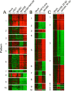
Fig. 1
Fig. 1. Effects of herpesvirus infection on host transcription
Wildtype (C57BL6/J) mice were infected with HSV1, MCMV, MHV68, latency-deficient MHV68 mutant (ORF73.stop), or both MCMV and MHV68 combined. All infections were performed with 1×10⁵ plaque forming units of virus, administered intranasally. Twenty-eight or 90 days post infection (dpi, as indicated), total RNA was purified from mouse spleens and host transcript levels assessed using Illumina Mouse Gene 6 (v1.1) arrays and raw data pre-normalized using Beadstudio software. The extraction of gene expression patterns and identification of genes through EPIG was performed using the microarray data (41, 42). Briefly, intensity values from all of the probe sets on the arrays were log2-transformed and adjusted by systematic variation normalization. Distinct patterns of gene expression were extracted on the basis of the expression profile correlation values, the minimum cluster size for the patterns, and the cluster-partitioning resolution. From the patterns and with a signal-to-noise ratio of 3 (p < 0.001), magnitude of 0.5 (1.4-fold change), and a correlation r value of 0.64 (p < 0.001) of the gene profiles, probe sets were selected. In this data set, the mock-infected group (28 dpi) was used as a reference state. The average of the replicates of mock group arrays was aligned to 0 as a baseline, with all other treated samples adjusted by the same amount. Shown are heatmaps from biological triplicate experiments with one mouse per group that identify transcriptional signatures correlating with different single or multiple herpesvirus infections (A, 28 dpi), acute vs. latent MHV68 infection (B, 28 dpi), or early (28 dpi) vs. late (90 dpi) latent MHV68 infection (C). EPIG identified gene lists are available in Supplementary Tables linked from the online version of this manuscript.
Fig. 2
Fig. 2. Latent MHV68 infection is associated with enhanced inflammatory cytokine production
C57BL/6 mice were infected with MHV68 at 1×10⁴ plaque forming units of virus, administered intranasally. Serum was harvested at the indicated time points (dpi) and assayed for cytokine concentrations via multiplex suspension array (RANTES, IL-6) or cytometric bead array (TNFα, IFNγ) according to the manufacturer's instructions (Bio Plex, Bio Rad Laboratories, Hercules, CA; BD Biosciences, San Diego, CA). Data are pooled from two to three independent experiments. Each dot represents one mouse. p values were calculated using a two-tailed Wilcoxon match pairs sign rank test, comparing mock to infected mice. *, p<0.05; **, p<0.01. Data for RANTES and IL-6, are unpublished results (Douglas W. White and Herbert W. Virgin). Data for TNFα and IFNγ are adapted from (48).
Fig. 3
Fig. 3. Hypothetical relationships between herpesvirus immune modulation during latency and cancer development
Shown are two models of cancer development that emphasize either genetic changes in the cancerous cell (Genocentric Model, A) or the immunologic microenvironment of the developing tumor (B) as driving forces in cancer progression. In genocentric models of cancer (A), the predominant view of herpesviruses (HV) is one of mutagenesis, either directly via introduction of viral oncogenes or indirectly. In immunologic models of cancer progression, HV may have more diverse mechanisms of interaction with the nascent cancer cell, including enhanced immunosurveillance, tumor elimination, or maintenance of pre-cancerous equilibrium triggered by latency-driven immune modulation. In contrast, the immune modulatory environment triggered by HV latency may promote more rapid tumor immunoediting and selection of immune evasion or suppressive tumor cells. In addition, compensatory regulatory events triggered by HV latency may enhance the development or recruitment of immunosuppressive cells into the tumor microenvironment. Hallmarks of cancer progression are shown the left in shaded triangles. Potential negative (tumor-promoting) mechanisms triggered by herpesvirus (HV) latency are indicated in red text with red arrows. Potential beneficial (tumor-inhibiting) mechanisms triggered by HV latency are indicated in green text with green arrows or blocked bars. The predominant modes of interaction between the immune system and cancerous cells are shown in blue text and based on (118).
Fig. 4
Fig. 4. Age-dependent outcomes in herpesvirus immunomodulation
(A). Herpesvirus latency as a mutualistic symbiosis to promote cross protection from secondary infection. This hypothesis postulates that all organisms are born with a basal level of innate resistance to infection. Specific pathogen-free mice represent this basal level. As humans acquire multiple herpesviruses during their early years of life, latency-driven immune responses trigger increased resistance to secondary infections. This cross protection may be transient or durable as described in the text. (B). If herpesviruses are acquired during early life, beneficial immune modulation including cross protection from infections and immune skewing away from Th2-driven allergic inflammation may result. In contrast, following herpesvirus infection later in life, latency-driven inflammation may exacerbate host inflammatory diseases. If infections are acquired predominantly in late childhood through adulthood, this model predicts that the primary outcomes for the host will be pathologic.

Similar articles

Cited by

References

    1. Pellett PE, Roizman B. The Family Herpesviridae: A Brief Introduction. In: Knipe DM, Howely PM, editors. Fields Virology. Philadelphia: Lippincott Williams & Wilkins; 2007. pp. 2479–2500.
    1. Roizman B, Knipe DM, Whitley RJ. Herpes Simplex Viruses. In: Knipe DM, Howely PM, editors. Fields Virology. Philadelphia: Lippincott Williams & Wilkins; 2007. pp. 2501–2602.
    1. Rickinson AB, Kieff E. Epstein-Barr Virus. In: Knipe DM, Howely PM, editors. Fields Virology. Philadelphia: Lippincott Williams & Wilkins; 2007. pp. 2655–2700.
    1. Ganem D. Kapsoi's Sarcoma-Associated Herpesvirus. In: Knipe DM, Howely PM, editors. Fields Virology. Philadelphia: Lippincott Williams & Wilkins; 2007. pp. 2847–2888.
    1. Mocarski ES, Shenk T, Pass RF. Cytomegaloviruses. In: Knipe DM, Howely PM, editors. Fields Virology. Philadelphia: Lippincott Williams & Wilkins; 2007. pp. 2701–2772.

Publication types

LinkOut - more resources