. 2011 Dec 14;13(1):80.
doi: 10.1186/1532-429X-13-80.

Cardiovascular magnetic resonance tagging of the right ventricular free wall for the assessment of long axis myocardial function in congenital heart disease

Affiliations

Cardiovascular magnetic resonance tagging of the right ventricular free wall for the assessment of long axis myocardial function in congenital heart disease

Sylvia S M Chen et al. J Cardiovasc Magn Reson. .

Abstract

Background: Right ventricular ejection fraction (RV-EF) has traditionally been used to measure and compare RV function serially over time, but may be a relatively insensitive marker of change in RV myocardial contractile function. We developed a cardiovascular magnetic resonance (CMR) tagging-based technique with a view to rapid and reproducible measurement of RV long axis function and applied it in patients with congenital heart disease.

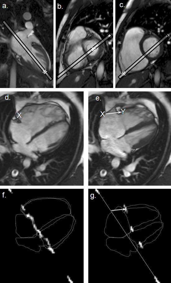
Methods: We studied 84 patients: 56 with repaired Tetralogy of Fallot (rTOF); 28 with atrial septal defect (ASD): 13 with and 15 without pulmonary hypertension (RV pressure > 40 mmHG by echocardiography). For comparison, 20 healthy controls were studied. CMR acquisitions included an anatomically defined four chamber cine followed by a cine gradient echo-planar sequence in the same plane with a labelling pre-pulse giving a tag line across the basal myocardium. RV tag displacement was measured with automated registration and tracking of the tag line together with standard measurement of RV-EF.

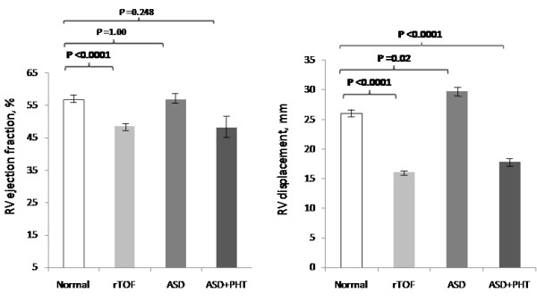
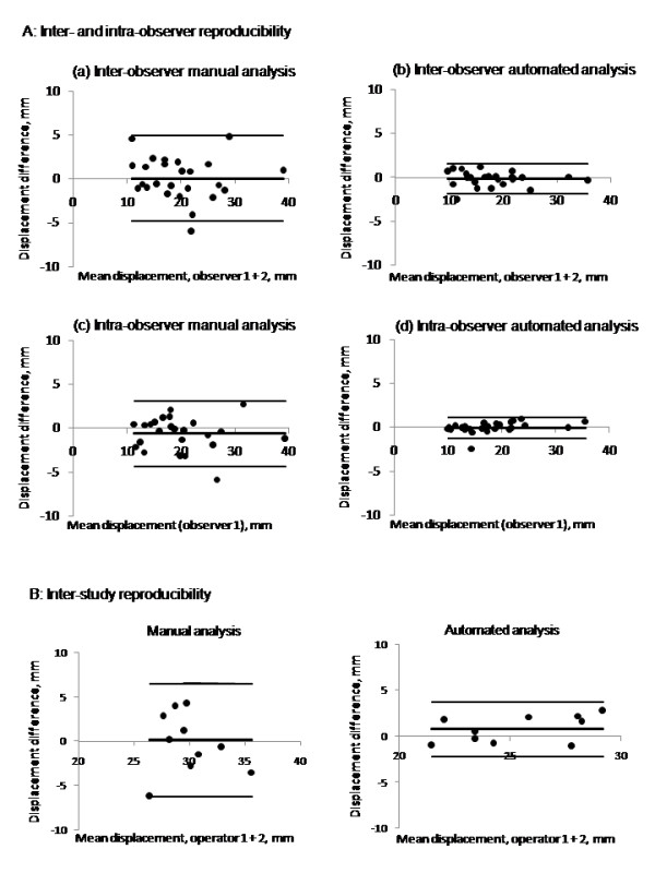
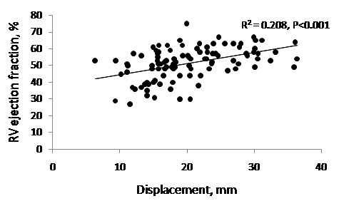
Results: Mean RV displacement was higher in the control (26 ± 3 mm) than in rTOF (16 ± 4 mm) and ASD with pulmonary hypertension (18 ± 3 mm) groups, but lower than in the ASD group without (30 ± 4 mm), P < 0.001. The technique was reproducible with inter-study bias ± 95% limits of agreement of 0.7 ± 2.7 mm. While RV-EF was lower in rTOF than in controls (49 ± 9% versus 57 ± 6%, P < 0.001), it did not differ between either ASD group and controls.

Conclusions: Measurements of RV long axis displacement by CMR tagging showed more differences between the groups studied than did RV-EF, and was reproducible, quick and easy to apply. Further work is needed to assess its potential use for the detection of longitudinal changes in RV myocardial function.


Figures

Figure 1
Figure 1
Alignment of the 4-chamber cine and the tagging by CMR. Three points marked (*) in the scout images (a-c) were used to define the 4-chamber image plane (d and e). The post-subtraction CMR tissue tag images at end-diastole (f) and end-systole (g) are shown superimposed on outlines traced from the corresponding 4-chamber cine frames. The white arrow in panel e indicates the systolic displacement of the RV free wall tag relative to a dotted line that has been drawn between the motionless tagged regions of the chest wall. For manual measurement of RV basal free wall displacement, by using CMRtools, a marker (X) was placed in the RV free wall 5 mm from the atrio-ventricular groove at end-diastole (e) and copied to the end-systolic frame (f), where the white arrow indicates the displacement measured (X→Y).
Figure 2
Figure 2
RV ejection fraction and RV displacement. Mean RV ejection fraction (± standard error) of the three patient groups compared to normal. RV ejection fraction in rTOF was significantly reduced compared to normal controls. There were no significant differences between the ASD groups and controls. Mean RV displacement (± standard error) in the three patient groups compared to normal. The rTOF and ASD with PHT groups were significantly lower compared to normal. In contrast, ASD without PHT were significantly higher than normal.
Figure 3
Figure 3
A and B: A (a-d). Inter- and intra-observer reproducibility of the manual and automated analyses. Bland-Altman plots showing inter-observer and intra-observer reproducibility of RV displacements by the manual (a and c) and automated (b and d) techniques. The automated analysis showed closer 95% limits of agreement than manual analysis. B. Inter-study reproducibility of RV displacement Bland-Altman plots showing inter-study reproducibility with closer 95% limits of agreement when analysed with the automated technique compared with the manual technique.
Figure 4
Figure 4
RV displacement and RV ejection fraction. There was a weak but significant correlation between RV displacement and RV ejection fraction.

References

    1. Therrien J, Provost Y, Merchant N, Williams W, Colman J, Webb G. Optimal timing for pulmonary valve replacement in adults after tetralogy of Fallot repair. Am J Cardiol. 2005;95:779–782. doi: 10.1016/j.amjcard.2004.11.037. - DOI - PubMed
    1. Sheehan F, Redington A. The right ventricle: anatomy, physiology and clinical imaging. Heart. 2008;94:1510–1515. doi: 10.1136/hrt.2007.132779. - DOI - PubMed
    1. Sehata ML, Cheng S, Osman NF, Bluemke DA, Lima JAC. Myocardial tissue tagging with cardiovascular magnetic resonance. J Cardiovasc Magn Reson. 2009;11:55. doi: 10.1186/1532-429X-11-55. - DOI - PMC - PubMed
    1. Haber I, Metaxas DN, Geva T, Axel L. Three-dimensional systolic kinematics of the right ventricle. Am J Physiol Heart Circ Physiol. 2005;289:1826–1833. doi: 10.1152/ajpheart.00442.2005. - DOI - PubMed
    1. Naito H, Arisawa J, Harada K, Yamagami H, Kozuka T, Tamura S. Assessment of right ventricular regional contraction and comparison with the left ventricle in normal humans: a cine magnetic resonance study with presaturation myocardial tagging. Br Heart J. 1995;74:186–191. doi: 10.1136/hrt.74.2.186. - DOI - PMC - PubMed

Publication types

MeSH terms