

Changes in human meibum lipid composition with age using nuclear magnetic resonance spectroscopy
- PMID: 22169100
- PMCID: PMC3292379
- DOI: 10.1167/iovs.11-8341
Changes in human meibum lipid composition with age using nuclear magnetic resonance spectroscopy
Abstract
Purpose: Human tear film stability decreases with increasing age. In this study, the changes in meibum composition were measured in search of markers of tear film instability.
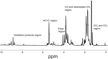
Methods: (1)H NMR nuclear magnetic resonance (NMR) spectra of 43 normal donors aged 1 to 88 years were acquired.
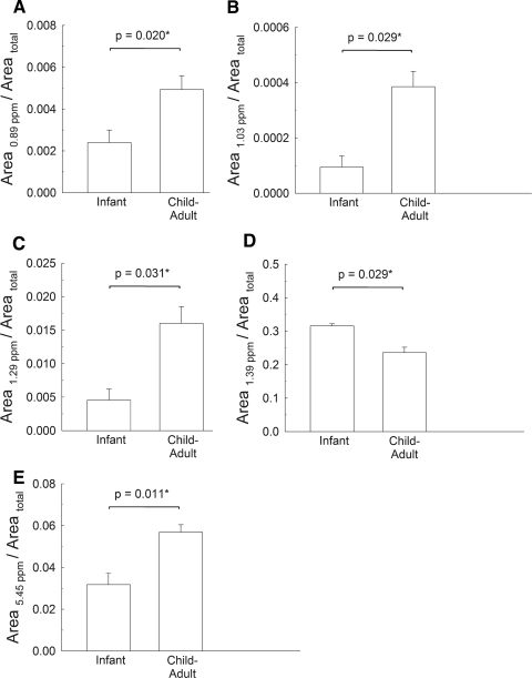
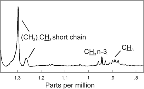
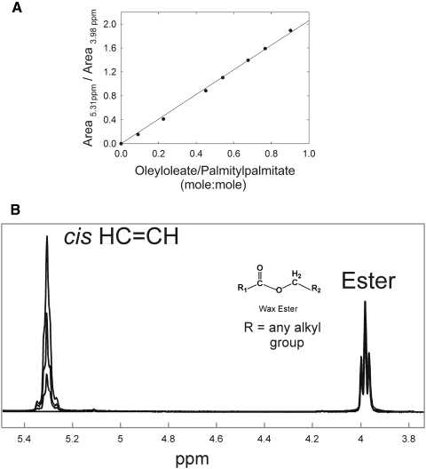
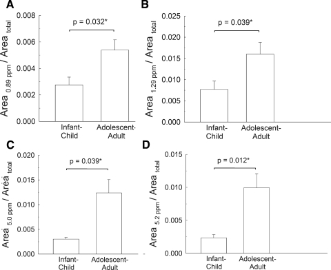
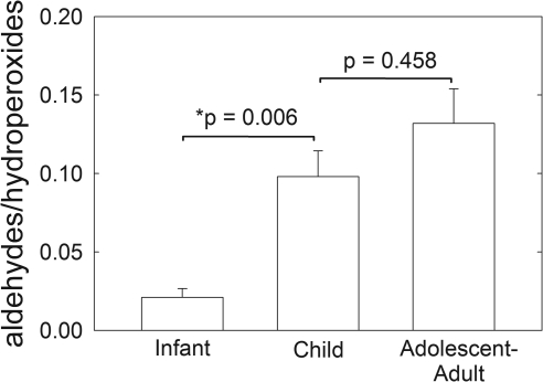
Results: Compared with meibum from adolescents and adults, meibum from infants and children contains less CH(3) and C═C groups and an increased aldehyde-to-lipid hydroperoxide ratio.
Conclusions: It is reasonable that tear film stability is higher in infants than in adults. Their meibum contains less CH(3) and C═C groups and higher levels of protein, and as a result, the lipid is more ordered because of the tighter and stronger lipid-lipid interactions. For water to evaporate, it must first pass through the tight lipid-lipid barrier. For tears to break up, lipid-lipid interactions must be broken. It is reasonable that because the lipid-lipid interactions are stronger in infants' and children's tears compared with those of adolescents and adults, the tear film in the younger groups is more stable and provides a better barrier to evaporation than does the tear film of adults. Lipid saturation could be the critical feature in meibum that stabilizes tears in infants.
Figures
Similar articles
-
Lipid order, saturation and surface property relationships: a study of human meibum saturation.Exp Eye Res. 2013 Nov;116:79-85. doi: 10.1016/j.exer.2013.08.012. Epub 2013 Aug 22. Exp Eye Res. 2013. PMID: 23973715
-
Physical changes in human meibum with age as measured by infrared spectroscopy.Ophthalmic Res. 2010;44(1):34-42. doi: 10.1159/000283606. Epub 2010 Feb 17. Ophthalmic Res. 2010. PMID: 20160464 Free PMC article.
-
Analysis of the composition of lipid in human meibum from normal infants, children, adolescents, adults, and adults with meibomian gland dysfunction using ¹H-NMR spectroscopy.Invest Ophthalmol Vis Sci. 2011 Sep 21;52(10):7350-8. doi: 10.1167/iovs.11-7391. Invest Ophthalmol Vis Sci. 2011. PMID: 21849420 Free PMC article.
-
The real reason for having a meibomian lipid layer covering the outer surface of the tear film - A review.Exp Eye Res. 2015 Aug;137:125-38. doi: 10.1016/j.exer.2015.05.002. Epub 2015 May 14. Exp Eye Res. 2015. PMID: 25981748 Review.
-
Analysis of meibum and tear lipids.Ocul Surf. 2012 Oct;10(4):230-50. doi: 10.1016/j.jtos.2012.07.004. Epub 2012 Jul 25. Ocul Surf. 2012. PMID: 23084145 Review.
Cited by
-
Asymptomatic Meibomian Gland Dysfunction and Cardiovascular Disease Risk Factors in a Middle-Aged Population in Taiwan - A Cross-sectional Analysis.Sci Rep. 2017 Jul 10;7(1):4935. doi: 10.1038/s41598-017-05368-z. Sci Rep. 2017. PMID: 28694455 Free PMC article.
-
Human Meibum Age, Lipid-Lipid Interactions and Lipid Saturation in Meibum from Infants.Int J Mol Sci. 2017 Aug 28;18(9):1862. doi: 10.3390/ijms18091862. Int J Mol Sci. 2017. PMID: 28846660 Free PMC article.
-
Effects of Aging on Human Meibum.Invest Ophthalmol Vis Sci. 2021 Sep 2;62(12):23. doi: 10.1167/iovs.62.12.23. Invest Ophthalmol Vis Sci. 2021. PMID: 34546321 Free PMC article.
-
Differences in human meibum lipid composition with meibomian gland dysfunction using NMR and principal component analysis.Invest Ophthalmol Vis Sci. 2012 Jan 25;53(1):337-47. doi: 10.1167/iovs.11-8551. Invest Ophthalmol Vis Sci. 2012. PMID: 22131391 Free PMC article.
-
Lipidomic analysis of human tear fluid reveals structure-specific lipid alterations in dry eye syndrome.J Lipid Res. 2014 Feb;55(2):299-306. doi: 10.1194/jlr.P041780. Epub 2013 Nov 28. J Lipid Res. 2014. PMID: 24287121 Free PMC article.
References
-
- Mantelli F, Tiberi E, Micera A, Lambiase A, Visintini F, Bonini S. MUC5AC over expression in tear film of neonates. Graefes Clin Exp Ophthalmol. 2007;245:1377–1381 - PubMed
-
- Isenberg SJ, Del Signore M, Chen A, Wei J, Guillon J. The lipid layer and stability of the preocular tear film in newborns and infants. Ophthalmology. 2003;110:1408–1411 - PubMed
-
- Sforza C, Rango M, Galante D, Bresolin N, Ferrario VF. Spontaneous blinking in healthy persons: an optoelectronic study of eyelid motion. OphthalmicPhysiol Opt. 2008;28:345–353 - PubMed
Publication types
MeSH terms
Substances
Grants and funding
LinkOut - more resources
Full Text Sources
Medical
