Review
. 2012 Feb;15(1):36-43.
doi: 10.1016/j.mib.2011.11.006. Epub 2011 Dec 12.

Pathogens and the placental fortress

Affiliations
Review

Pathogens and the placental fortress

Jennifer R Robbins et al. Curr Opin Microbiol. 2012 Feb.

Abstract

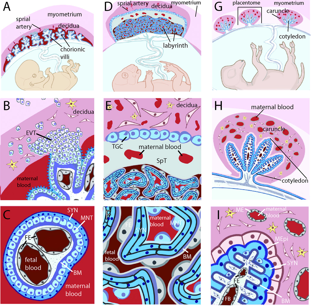
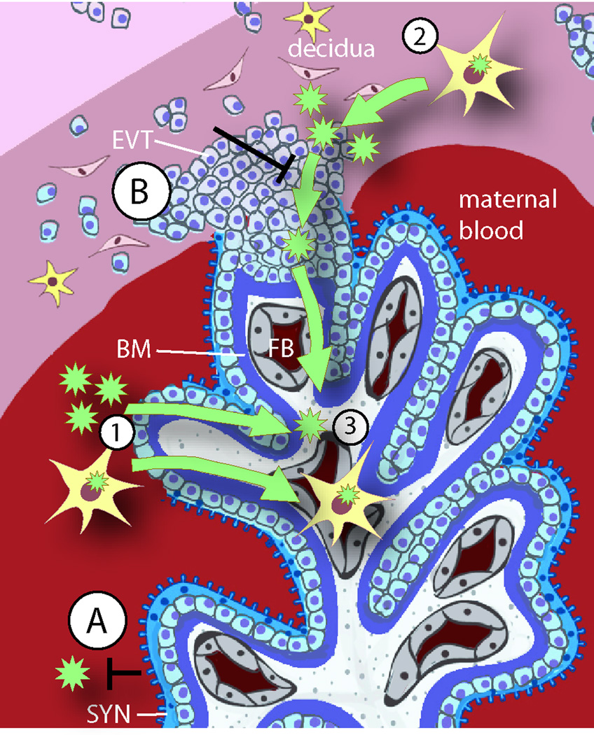
Placental infections are major causes of maternal and fetal disease. This review introduces a new paradigm for placental infections based on current knowledge of placental defenses and how this barrier can be breached. Transmission of pathogens from mother to fetus can occur at two sites of direct contact between maternal cells and specialized fetal cells (trophoblasts) in the human placenta: firstly, maternal immune and endothelial cells juxtaposed to extravillous trophoblasts in the uterine implantation site and secondly, maternal blood surrounding the syncytiotrophoblast (SYN). Recent findings suggest that the primary vulnerability is in the implantation site. We explore evidence that the placental SYN evolved as a defense against pathogens, and that inflammation-mediated spontaneous abortion may benefit mother and pathogen.


Figures

Fig. 1
Fig. 1. Placental structures are diverse and do not necessarily cluster evolutionarily, complicating the selection of appropriate model systems
A. The hemomonochorial human placenta is a villous structure bathed in maternal blood and anchored deep in the uterine decidua by extravillous trophoblasts. B. The human uterine-trophoblast interface is composed of decidual fibroblasts and maternal leukocytes juxtaposed with fetal extravillous trophoblasts (EVT), some of which migrate into the myometrium, while others remodel maternal spiral arteries (not shown). Many remain connected to a column of trophoblasts that extends into the fetal villus and stops at a basement membrane that divides trophoblasts from fetal capillaries. C. The human blood-trophoblast interface, showing villus cross-section. Maternal blood is separated from fetal vessels by a single syncytiotrophoblast (SYN), whose apical surface is covered by dense branched microvilli. Underlying that is a layer of mononuclear subsyncytial trophoblasts (MNT) that is continuous in early pregnancy but semi-discontinuous in later trimesters. Both structures are undergirded by a basement membrane. D. The hemotrichorial mouse placenta places maternal and fetal vessels in close contact within a nutrient-exchange labyrinth that is anchored in the decidua E. via a spongiotrophoblast (SpT) region and trophoblast giant cells (TGC) (analogous to EVT). The decidua is primarily composed of leukocytes and fibroblasts, as well as maternal arteries modified by trophoblast giant cells (not shown). F. Cross-section of labyrinthine region. From the maternal blood, a semi-discontinuous layer of mononuclear trophoblasts (MNT) overlays two layers of synctiotrophoblast with limited, non-overlapping cell-cell junctions. Beyond this lies fetal endothelium (FE). G. The sheep placenta is primarily epitheliochorial, and composed of multiple placentomes throughout the uterus. H. Each placentome includes a uterine caruncle rich in endometrial glands (not shown) and partially encapsulating a relatively non-invasive fetal cotyledon. I. Much of the cotyledonary surface effaces with a maternal epithelium (MEpi; which is not fully continuous and considered synepitheliochorial in regions, not shown). Maternal blood is separated from fetal blood by maternal endothelium (ME), endometrial fibroblasts, maternal epithelium, fetal syncytiotrophoblast (SYN), chorionic fibroblasts and fetal endothelium (FE). B–I. yellow cells = maternal leukocytes. A-F. Adapted from [5].
Fig. 2
Fig. 2. Placental colonization by pathogens
A schematic depiction of the human maternal-fetal interface is used to illustrate the barriers and possible pathways for pathogen transmission. The two sites of direct contact between maternal and fetal cells are the blood-syncytiotrophoblast (SYN) (A) and the uterus-extravillous trophoblast (EVT) (B) interfaces. Both of these trophoblast subpopulations have defense mechanisms against infection, and underneath the trophoblast barrier, the basement membrane (BM) presents an additional physical barrier. Failure of the placental barrier can occur in the presence of high pathogen titers or multiple infections. Damage of the syncytiotrophoblast enables pathogens (green stars) that are free in maternal blood or inside of maternal leukocytes (yellow cells with green stars) to cross into fetal tissues (1). However, animal and organ culture models agree that most placental infections originate in the uterine decidua (2), which is minimally accessible from the maternal blood. Pathogens can reach the decidua only by dissemination in maternal cells, most likely leukocytes. If the defense mechanisms of the EVT are overcome, the infection may spread to the fetal blood (FB) (3), act as a nidus for maternal reinfection, and/or cause trophoblast death resulting in placental insufficiency or spontaneous abortion. Finally, some pathogens may reach the fetus by traveling within maternal leukocytes on their natural way to the fetus.

References

    1. Beck S, Wojdyla D, Say L, Betran AP, Merialdi M, Requejo JH, Rubens C, Menon R, Van Look PF. The worldwide incidence of preterm birth: A systematic review of maternal mortality and morbidity. Bull World Health Organ. 2010;88(1):31–38. - PMC - PubMed
    1. Goldenberg RL, Culhane JF, Iams JD, Romero R. Epidemiology and causes of preterm birth. Lancet. 2008;371(9606):75–84. - PMC - PubMed
    1. Saigal S, Doyle LW. An overview of mortality and sequelae of preterm birth from infancy to adulthood. Lancet. 2008;371(9608):261–269. - PubMed
    1. Givens MD, Marley MS. Infectious causes of embryonic and fetal mortality. Theriogenology. 2008;70(3):270–285. - PMC - PubMed
    1. Maltepe E, Bakardjiev AI, Fisher SJ. The placenta: Transcriptional, epigenetic, and physiological integration during development. J Clin Invest. 2010;120(4):1016–1025. - PMC - PubMed

Publication types

MeSH terms