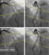
In vivo assessment of bifurcation optimal viewing angles and bifurcation angles by three-dimensional (3D) quantitative coronary angiography
- PMID: 22169957
- PMCID: PMC3473185
- DOI: 10.1007/s10554-011-9996-x
In vivo assessment of bifurcation optimal viewing angles and bifurcation angles by three-dimensional (3D) quantitative coronary angiography
Abstract
Evaluation and stenting of coronary bifurcation lesions may benefit from optimal angiographic views. The anatomy-defined bifurcation optimal viewing angle (ABOVA) is characterized by having an orthogonal view of the bifurcation, such that overlap and foreshortening at the ostium are minimized. However, due to the mechanical constraints of the X-ray systems, certain deep angles cannot be reached by the C-arm. Therefore, second best or, so-called obtainable bifurcation optimal viewing angle (OBOVA) has to be used as an alternative. This study assessed the distributions of ABOVA and OBOVA using 3D quantitative coronary angiography in a typical patient population. In addition, the bifurcation angles in four main coronary bifurcations were assessed and compared. Patients with obstructive coronary bifurcation disease were included in this multicenter registry. A novel and validated 3D QCA software package was applied to reconstruct the bifurcations and to calculate the bifurcation angles in 3D. A list of optimal viewing angle candidates including ABOVA was also automatically proposed by the software. In a next step, the operator selected the best viewing angle as OBOVA, while applying a novel overlap prediction approach to assure no overlap between the target bifurcation and other major coronary arteries. A total of 194 bifurcations from 181 patients were assessed. The ABOVA could not be reached in 56.7% of the cases; being 40 (81.6%), 40 (78.4%), 9 (17.6%), and 21 (48.8%) cases for LM/LAD/LCx, LAD/Diagonal, LCx/OM, and PDA/PLA, respectively. Both ABOVA and OBOVA distributed sparsely with large ranges of variance: LM/LAD/LCx, 5 ± 33 RAO, 47 ± 35 Caudal versus 4 ± 39 LAO, 35 ± 16 Caudal; LAD/Diagonal, 4 ± 38 RAO, 50 ± 14 Cranial versus 14 ± 28 LAO, 33 ± 5 Cranial; LCx/OM, 21 ± 32 LAO, 27 ± 17 Caudal versus 18 ± 31 LAO, 25 ± 13 Caudal; PDA/PLA, 34 ± 21 LAO, 36 ± 21 Cranial versus 28 ± 25 LAO, 29 ± 15 Cranial. LM/LAD/LCx had the smallest proximal bifurcation angle (128° ± 24°) and the largest distal bifurcation angle (80° ± 21°), as compared with LAD/Diagonal (151° ± 13º and 48° ± 16º), LCx/OM (146° ± 18º and 57° ± 16°), and PDA/PLA (145° ± 19° and 59° ± 17°). In conclusion, large variabilities in optimal viewing angles existed for all main coronary bifurcations. The anatomy-defined bifurcation optimal viewing angle could not be reached in vivo in roughly half of the cases due to the mechanical constraints of the current X-ray systems. Obtainable bifurcation optimal viewing angle should be provided as an alternative or second best. The bifurcation angles in the left main bifurcation demonstrated the largest variabilities.
Figures


References
-
- Suzuki N, Angiolillo DJ, Kawaguchi R, Futamatsu H, Bass TA, Costa MA. Percutaneous coronary intervention of bifurcation coronary disease. Minerva Cardioangiol. 2007;55:57–71. - PubMed
-
- Lemos PA, Saia F, Ligthart JMR, Arampatzis CA, Sianos G, Tanabe K, et al. Coronary restenosis after sirolimus-eluting stent implantation: morphological description and mechanistic analysis from a consecutive series of cases. Circulation. 2003;108:257–260. doi: 10.1161/01.CIR.0000083366.33686.11. - DOI - PubMed
Publication types
MeSH terms
LinkOut - more resources
Full Text Sources
Other Literature Sources
Medical

