. 2012 Feb 3;287(6):4248-59.
doi: 10.1074/jbc.M111.292243. Epub 2011 Dec 13.

Intersubunit bridge formation governs agonist efficacy at nicotinic acetylcholine α4β2 receptors: unique role of halogen bonding revealed

Affiliations

Intersubunit bridge formation governs agonist efficacy at nicotinic acetylcholine α4β2 receptors: unique role of halogen bonding revealed

Line Aagot Hede Rohde et al. J Biol Chem. .

Abstract

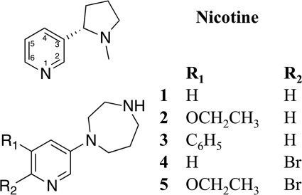
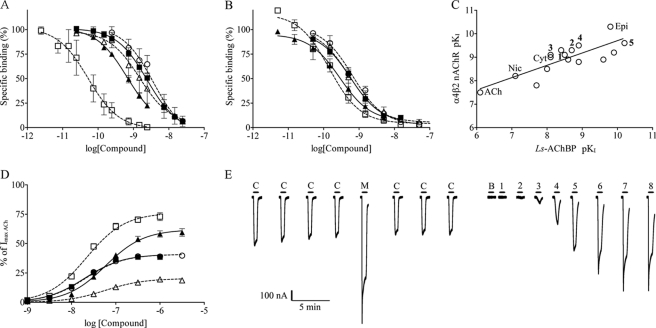
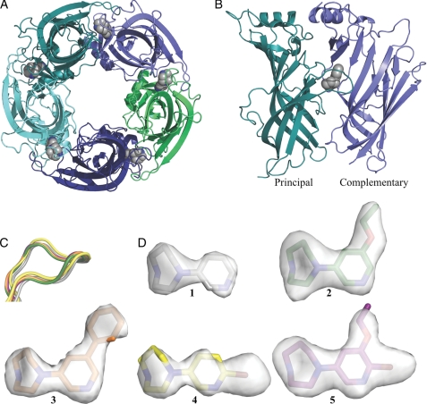
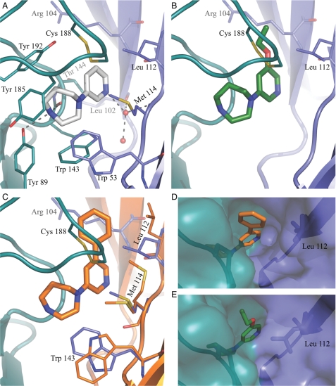
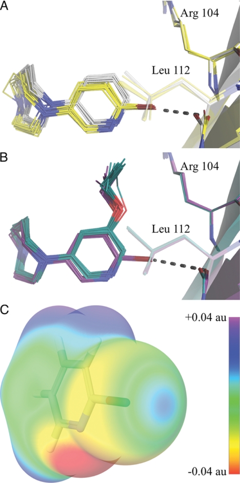
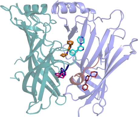
The α4β2 subtype of the nicotinic acetylcholine receptor has been pursued as a drug target for treatment of psychiatric and neurodegenerative disorders and smoking cessation aids for decades. Still, a thorough understanding of structure-function relationships of α4β2 agonists is lacking. Using binding experiments, electrophysiology and x-ray crystallography we have investigated a consecutive series of five prototypical pyridine-containing agonists derived from 1-(pyridin-3-yl)-1,4-diazepane. A correlation between binding affinities at α4β2 and the acetylcholine-binding protein from Lymnaea stagnalis (Ls-AChBP) confirms Ls-AChBP as structural surrogate for α4β2 receptors. Crystal structures of five agonists with efficacies at α4β2 from 21-76% were determined in complex with Ls-AChBP. No variation in closure of loop C is observed despite large efficacy variations. Instead, the efficacy of a compound appears tightly coupled to its ability to form a strong intersubunit bridge linking the primary and complementary binding interfaces. For the tested agonists, a specific halogen bond was observed to play a large role in establishing such strong intersubunit anchoring.


Figures

FIGURE 1.
FIGURE 1.
Structures of nicotine and compounds 1-5.
FIGURE 2.
FIGURE 2.
Binding and functional profile of compounds 1–5: 1, ○; 2, ■; 3, △; 4, ▴; 5, □. A, binding affinity (±S.E.) at Ls-AChBP ([3H]epibatidine) is shown. B, binding affinity (±S.E.) at α4β2 nAChR ([3H]cytisine) is shown. C, correlation between binding affinities of agonists at Ls-AChBP and α4β2 nAChR is shown. Slope = 0.5, R2 = 0.7. Nic, nicotine; epi, epibatidine; Cyt, cytisine; unnumbered compounds correspond to compounds listed in supplemental Table S1. D, functional concentration-response profiles for 1–5 from HS α4β2 nAChRs are expressed from the dimeric concatenated β2–6-α4 construct and β2 in a 4:1 ratio (±S.E.). Responses are relative to a maximum response of ACh. E, representative current traces for compound 5 in two-electrode voltage-clamp electrophysiological experiments in X. laevis oocytes. Oocyte were injected with β2–6-α4 and β2 nAChR subunits in a 4:1 ratio. Application of the compound is indicated by a bar above each trace, and C denotes an ACh control concentration of 1 μm, M denotes a 100 μm AChmax concentration, B denotes a buffer application; numbers 1–8 denote increasing concentrations of compound 5 in half-log-unit increments with a minimal concentration of 316 pm in application, 1, and maximal concentration of 1 μm in application 8.
FIGURE 3.
FIGURE 3.
Structure of Ls-AChBP co-crystallized with partial agonists. A, shown is a bottom view of the pentameric Ls-AChBP with compound 1 (gray) in the orthosteric agonist binding pockets. Subunits are colored individually. B, shown is the Ls-AChBP interface with compound 1 (gray) bound under the C-loop. The principal and complementary sides are in teal and blue, respectively. C, the C-loop of AChBP in complex with compound 1 (gray), 2 (green), 3 (orange), 4 (yellow) and 5 (purple) is shown. The structures were superimposed on the principal subunit. D, the conformations of compounds 1–5 when bound in the binding pocket of Ls-AChBP. FoFc omit maps are shown and contoured at 3.0δ.
FIGURE 4.
FIGURE 4.
Detailed binding modes for compounds 1–3. A, shown is the binding mode of compounds 1 (gray) in Ls-AChBP. Residues interacting with 1 are colored teal at the principal side and blue at the complementary side. Broken lines indicate ionic interactions with the protein or water molecule (red sphere) within 3.5 Å. B, the binding mode of compounds 2 (green) is shown. Side chains interacting with the ethoxy substituent are shown. C, the binding mode of compound 3 (orange) is shown. Orange residues (complementary side) are shown. Blue residues from the 1-AChBP complex are included to illustrate the conformational changes in the protein associated with binding of 3. D and E, shown is the surface representation of the 3-AChBP and 2-AChBP complexes illustrating the broad (D) and narrow (E) channel to the surface of the protein.
FIGURE 5.
FIGURE 5.
Halogen bonding and binding mode of halogen-substituted compounds 4 and 5. All binding sites are superimposed as described under “Experimental Procedures.” A, shown is a comparison of the binding modes of compounds 1 (gray carbons) and 4 (yellow carbons). B, shown is a comparison of the binding modes of compounds 2 (green carbons) and 5 (purple carbons). The broken line indicates halogen bond. C, shown is the electrostatic potential of 2-bromopyridine mapped on the surface of the molecular electron density calculated using the B3LYP hybrid potential and a cc-pvdzs basis set. au, atomic units.
FIGURE 6.
FIGURE 6.
Collective image of additional binding orientations observed for compound 3. Superimposition of A/B and H/I interfaces. The principal side is colored teal, and the complementary side is blue. Compound 3 in the orthosteric binding site as it is found in all interfaces is colored orange, and 3 in the H/I and A/B interfaces are bluish (dark blue and cyan) and reddish (pink and red), respectively. The molecule of 3 binding to the β-sheets sandwich on the complementary side induces an ordered α-helical turn in loop-F (also colored red).

Similar articles

Cited by

References

    1. Schneider J. S., Van Velson M., Menzaghi F., Lloyd G. K. (1998) Effects of the nicotinic acetylcholine receptor agonist SIB-1508Y on object retrieval performance in MPTP-treated monkeys. Comparison with levodopa treatment. Ann. Neurol. 43, 311–317 - PubMed
    1. Levin E. D., Christopher N. C., Weaver T., Moore J., Brucato F. (1999) Ventral hippocampal ibotenic acid lesions block chronic nicotine-induced spatial working memory improvement in rats. Brain Res. Cogn. Brain Res. 7, 405–410 - PubMed
    1. Steinlein O. K., Mulley J. C., Propping P., Wallace R. H., Phillips H. A., Sutherland G. R., Scheffer I. E., Berkovic S. F. (1995) A missense mutation in the neuronal nicotinic acetylcholine receptor α4 subunit is associated with autosomal dominant nocturnal frontal lobe epilepsy. Nat. Genet. 11, 201–203 - PubMed
    1. Grottick A. J., Higgins G. A. (2000) Effect of subtype selective nicotinic compounds on attention as assessed by the five-choice serial reaction time task. Behav Brain Res. 117, 197–208 - PubMed
    1. Jensen A. A., Frølund B., Liljefors T., Krogsgaard-Larsen P. (2005) Neuronal nicotinic acetylcholine receptors. Structural revelations, target identifications, and therapeutic inspirations. J. Med. Chem. 48, 4705–4745 - PubMed

LinkOut - more resources