. 2012 Feb 3;287(6):4311-22.
doi: 10.1074/jbc.M111.310300. Epub 2011 Dec 14.

Calcium-induced folding and stabilization of the Pseudomonas aeruginosa alkaline protease

Affiliations

Calcium-induced folding and stabilization of the Pseudomonas aeruginosa alkaline protease

Liang Zhang et al. J Biol Chem. .

Abstract

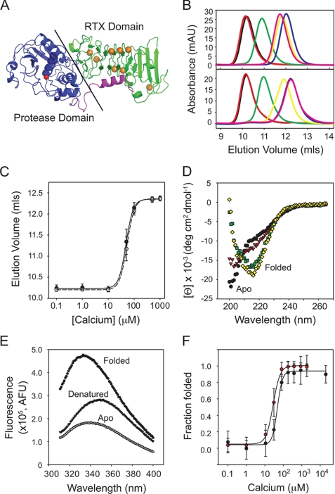
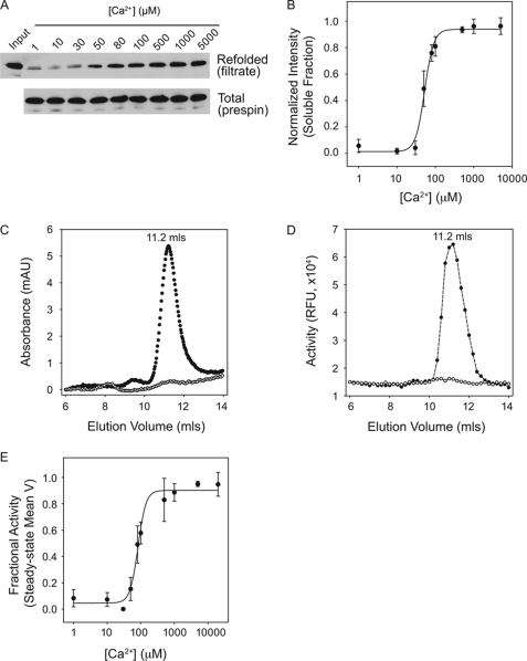
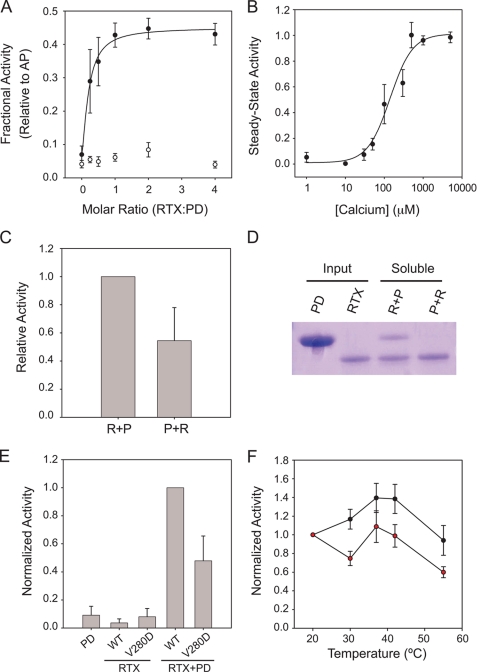
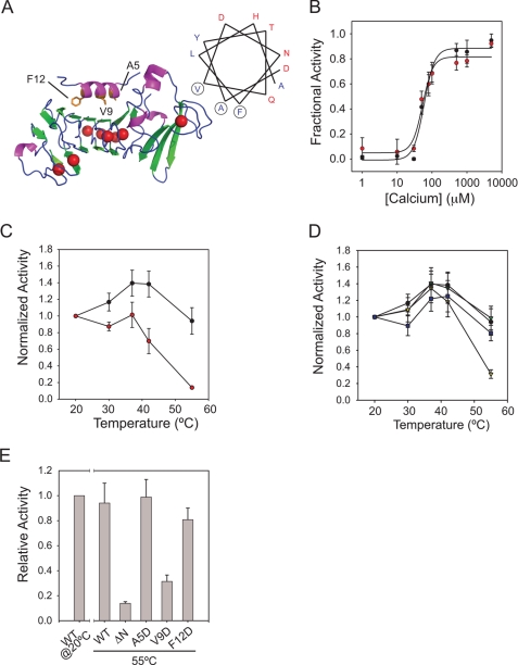
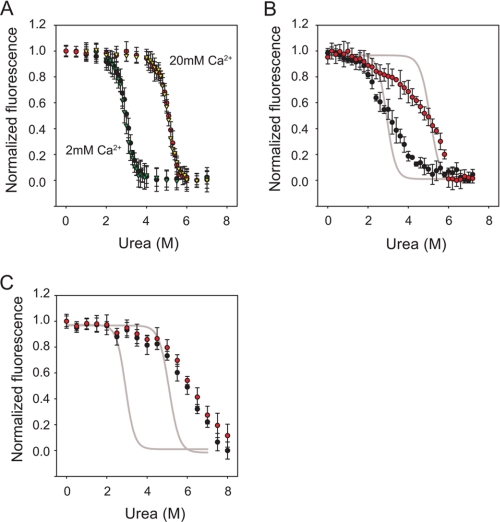
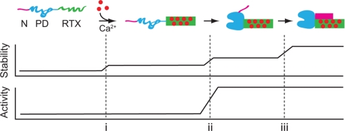
Pseudomonas aeruginosa is an opportunistic pathogen that contributes to the mortality of immunocompromised individuals and patients with cystic fibrosis. Pseudomonas infection presents clinical challenges due to its ability to form biofilms and modulate host-pathogen interactions through the secretion of virulence factors. The calcium-regulated alkaline protease (AP), a member of the repeats in toxin (RTX) family of proteins, is implicated in multiple modes of infection. A series of full-length and truncation mutants were purified for structural and functional studies to evaluate the role of Ca(2+) in AP folding and activation. We find that Ca(2+) binding induces RTX folding, which serves to chaperone the folding of the protease domain. Subsequent association of the RTX domain with an N-terminal α-helix stabilizes AP. These results provide a basis for the Ca(2+)-mediated regulation of AP and suggest mechanisms by which Ca(2+) regulates the RTX family of virulence factors.


Figures

FIGURE 1.
FIGURE 1.
Calcium-induced folding of the alkaline protease RTX domain. The isolated RTX domain from alkaline protease was purified for in vitro Ca2+ binding and structural studies. A, a schematic representation of the 1KAP crystal structure is shown (36). The Ca2+-binding RTX domain is shown in green, the protease domain is shown in blue, and the N-terminal helix is shown in magenta. The active site Zn2+ is shown as a red sphere, and the RTX-bound Ca2+ ions are shown in orange. B, representative analytical gel filtration chromatographs show the changes in Ca2+-induced hydrodynamic radii. Calcium-induced protein folding was evaluated by injecting Ca2+-free protein onto a column pre-equilibrated with increasing [Ca2+] (top). Bottom, unfolding was monitored by injecting Ca2+-bound protein onto the column with decreasing [Ca2+]. Calcium concentrations were 1 μm (black), 10 μm (red), 50 μm (green), 100 μm (yellow), 500 μm (magenta), and 1 mm (blue). C, a summary of the Ca2+-induced hydrodynamic changes is shown and fit using non-linear regression. The calcium-induced folding transition is shown by the solid circles and line. The unfolding transition is shown in open circles and dashed lines. D, representative circular dichroism spectra of the Ca2+-induced changes in RTX secondary structure are shown. Calcium concentrations were 0 μm (black circles), 20 μm (red triangles), 50 μm (green squares), and 200 μm (yellow diamonds). E, representative fluorescence spectra are shown for the apo state (filled circles), Ca2+-bound folded state (open circles), and urea-induced denatured state (solid triangles). F, summary of the folding transitions monitored by CD (black circles) and fluorescence (red circles). Data are shown as mean ± S.D. (error bars).
FIGURE 2.
FIGURE 2.
Calcium-induced AP folding. Full-length alkaline protease was purified for binding and activation studies to evaluate the role of Ca2+ binding on AP folding. A, AP refolding was assessed after rapid dilution into Ca2+-containing buffers and filtration through a Microcon YM-100 filter. A representative Western blot of the filtrate (top) and total protein (bottom) is shown for Ca2+ concentrations between 1 μm and 5 mm. B, the refolded fraction of AP after filtration was assessed by densitometry and is shown as a function of Ca2+ in the refolding buffer. C, analytical gel filtration chromatography was used to evaluate the hydrodynamic radius of purified AP. Protein was refolded in the absence of Ca2+ (open circles) or in 20 mm Ca2+ (filled circles) and filtered prior to column injection. D, protease activity was assessed from fractions collected after gel filtration. Proteins refolded in the absence of Ca2+ (open circles) and in 20 mm Ca2+ (filled circles) were evaluated utilizing the BODIPY-casein substrate. E, mean velocities of the steady-state activities of AP refolded are shown plotted against Ca2+. Non-linear, least squares regression fit to a four-parameter Hill equation is shown as a solid line in B and E. Data are shown as mean ± S.D. (error bars).
FIGURE 3.
FIGURE 3.
Calcium-regulated interactions of the RTX and PD domains. To assess the coupling of RTX folding and PD folding and activation, the interaction between these two domains was evaluated by trans-complementation studies and mutagenesis. A, trans-activation of the PD by refolded RTX is shown as a function of the molar ratio between the RTX and PD domains. PD was diluted into buffers containing refolded RTX in 2 mm Ca2+ (filled circles) or in the absence of Ca2+ (open circles). Data are normalized to the activity of the full-length AP protein refolded under identical conditions. The non-linear regression fit is shown. B, a summary of Ca2+ titration with a 2:1 ratio of RTX to PD is shown in filled circles and fit using non-linear regression. As in A, the RTX domain was refolded prior to dilution of the PD. C, order of addition studies were used to assess the potential role of RTX in the folding or activation of PD. Refolding the RTX prior to the PD (R + P) resulted in a nearly 2-fold increase in protease activity when compared with the activity seen when the PD was refolded and then activated by the addition of RTX (P + R). D, a representative Coomassie Blue-stained gel of refolded RTX and PD proteins from the trans-activation studies is shown. The input protein is shown for both the PD and RTX proteins. Refolding the PD in the presence of the folded RTX (R + P) results in solubilization of the PD after separation by centrifugation. The addition of the RTX domain to PD refolded alone resulted in significantly reduced recovery of the PD after centrifugation, demonstrating that folding of PD was dependent on the presence of the folded RTX. E, disruption of the RTX-PD interaction results in decreased trans-activation. A summary of protease activities of the PD, the WT and V280D RTX domains, and mixtures containing the PD with the RTX domains is shown after refolding in 2 mm Ca2+. The V280D substitution, in the core of the RTX-PD interface, decreases trans-activation of the PD by ∼50% as compared with WT, consistent with a disruption of the appropriate RTX-PD association. F, full-length AP is sensitive to the introduction of the V280D mutation. Protease activity as a function of reaction temperature is shown for the WT (black circles) and V280D (red circles) proteins using the casein digestion assay. The V280D mutant refolds with an efficiency similar to that of wild type but shows an increased thermal sensitivity at all elevated reaction temperatures. Data shown are mean ± S.D. (error bars).
FIGURE 4.
FIGURE 4.
N-terminal helix stabilization of AP. The role of the N-terminal helix in AP folding and stabilization was assessed using functional assays. A, schematic diagram of the RTX and N-terminal helix sequences of AP is shown with a helical wheel projection of the N-terminal helix. The locations of the A5D, V9D, and F12D missense mutations are indicated in the structure and circled in the helical wheel. B, calcium-induced refolding and activation were assessed for the wild type (black circles) and ΔN-AP proteins (red circles). Activities of the wild type and ΔN-AP proteins are normalized to their respective maxima in saturating Ca2+. C, the temperature sensitivities of AP (black circles) and ΔN-AP (red circles) activities are shown. D, the temperature sensitivity of the N-terminal helix missense mutations is shown as follows: wild type (black circles), A5D (purple squares), V9D (yellow triangles), and F12D (green triangles). E, summary data for the wild type, ΔN-AP, and N-terminal helix missense mutants are shown at 55 °C. Activity of the wild type protein at 20 °C is shown for reference. Data are presented as mean ± S.D. (error bars).
FIGURE 5.
FIGURE 5.
Urea-induced unfolding of RTX, ΔN-AP, and AP proteins. Urea denaturation titrations were performed to assess the relative stabilities of the RTX and AP proteins. A, RTX proteins in 2 and 20 mm Ca2+ were assessed by monitoring changes in tryptophan fluorescence. Folding/unfolding transitions were reversible in both 2 and 20 mm Ca2+ and are shown as follows: unfolding, 2 mm Ca2+ (black circles); refolding, 2 mm Ca2+ (green triangles); unfolding, 20 mm Ca2+ (red circles); refolding, 20 mm Ca2+ (yellow triangles). B, unfolding of the ΔN-AP proteins at 2 mm Ca2+ (black circles) and 20 mm Ca2+ (red circles) is shown. Sigmoidal fits of the RTX unfolding transitions at 2 and 20 mm Ca2+ are shown in gray lines for reference. The increase in apparent stability as a function of increasing [Ca2+] from 2 to 20 mm was similar for both the RTX and ΔN-AP proteins. C, unfolding of the full-length AP is shown at 2 mm Ca2+ (black circles) and 20 mm Ca2+ (red circles). The RTX unfolding transition is shown as a gray line, as in B. Data are presented as mean ± S.D. (error bars).
FIGURE 6.
FIGURE 6.
Alkaline protease folding pathway. A putative pathway for the vectoral folding and stabilization of AP is presented. Ca2+ binding to the RTX domain initiates its folding (i). The native RTX domain facilitates the folding and activation of the PD via the 1000-Å2 RTX-PD interface (ii). The folded PD, in turn, facilitates the N-terminal helix-RTX interaction by placing the N-terminal helix in position to bind the folded RTX domain (iii). Stability of AP increases with each domain-domain association. Stabilization of the RTX domain structure occurs upon its interaction with Ca2+, its association with the PD, and its association with the N-terminal helix. The activity of AP is regulated by the association of the PD with the folded, Ca2+-bound RTX domain. The AP domains are colored as follows: N-terminal helix (magenta), PD (blue), and RTX (green) as in Fig. 1A. Folded protein is shown as a solid shape; unfolded protein domains are shown as extended lines.

Similar articles

Cited by

References

    1. Lyczak J. B., Cannon C. L., Pier G. B. (2000) Establishment of Pseudomonas aeruginosa infection. Lessons from a versatile opportunist. Microbes Infect. 2, 1051–1060 - PubMed
    1. Hobden J. A. (2002) Pseudomonas aeruginosa proteases and corneal virulence. DNA Cell Biol. 21, 391–396 - PubMed
    1. Lyczak J. B., Cannon C. L., Pier G. B. (2002) Lung infections associated with cystic fibrosis. Clin. Microbiol. Rev. 15, 194–222 - PMC - PubMed
    1. Leidal K. G., Munson K. L., Johnson M. C., Denning G. M. (2003) Metalloproteases from Pseudomonas aeruginosa degrade human RANTES, MCP-1, and ENA-78. J. Interferon Cytokine Res. 23, 307–318 - PubMed
    1. Kharazmi A., Høiby N., Döring G., Valerius N. H. (1984) Pseudomonas aeruginosa exoproteases inhibit human neutrophil chemiluminescence. Infect. Immun. 44, 587–591 - PMC - PubMed

Publication types

MeSH terms

LinkOut - more resources