. 2012 Feb 3;287(6):4335-47.
doi: 10.1074/jbc.M111.314831. Epub 2011 Dec 14.

Fucose-binding lectin from opportunistic pathogen Burkholderia ambifaria binds to both plant and human oligosaccharidic epitopes

Affiliations

Fucose-binding lectin from opportunistic pathogen Burkholderia ambifaria binds to both plant and human oligosaccharidic epitopes

Aymeric Audfray et al. J Biol Chem. .

Abstract

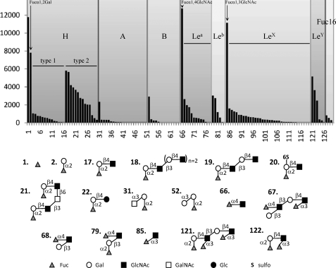
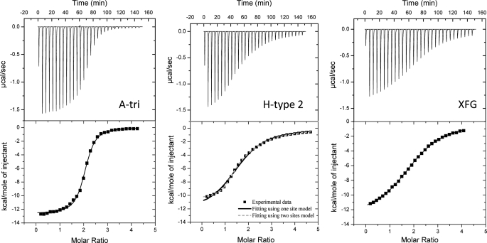
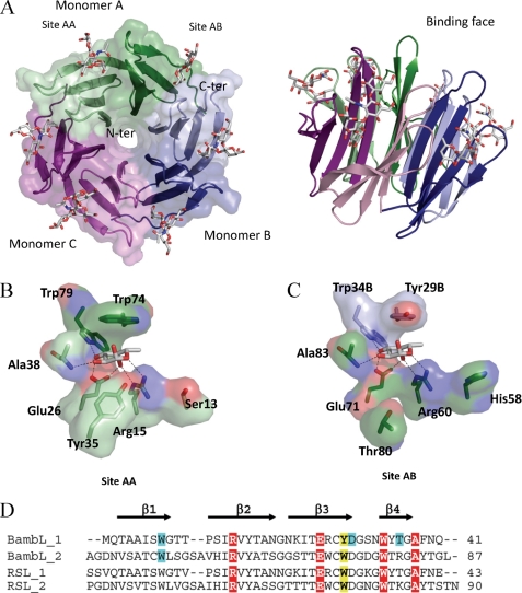
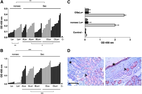
Burkholderia ambifaria is generally associated with the rhizosphere of plants where it has biocontrol effects on other microorganisms. It is also a member of the Burkholderia cepacia complex, a group of closely related bacteria that cause lung infections in immunocompromised patients as well as in patients with granulomatous disease or cystic fibrosis. Our previous work indicated that fucose on human epithelia is a frequent target for lectins and adhesins of lung pathogens (Sulák, O., Cioci, G., Lameignère, E., Balloy, V., Round, A., Gutsche, I., Malinovská, L., Chignard, M., Kosma, P., Aubert, D. F., Marolda, C. L., Valvano, M. A., Wimmerová, M., and Imberty, A. (2011) PLoS Pathog. 7, e1002238). Analysis of the B. ambifaria genome identified BambL as a putative fucose-binding lectin. The 87-amino acid protein was produced recombinantly and demonstrated to bind to fucosylated oligosaccharides with a preference for αFuc1-2Gal epitopes. Crystal structures revealed that it associates as a trimer with two fucose-binding sites per monomer. The overall fold is a six-bladed β-propeller formed by oligomerization as in the Ralstonia solanacearum lectin and not by sequential domains like the fungal fucose lectin from Aleuria aurantia. The affinity of BambL for small fucosylated glycans is very high as demonstrated by microcalorimetry (K(D) < 1 μM). Plant cell wall oligosaccharides and human histo-blood group oligosaccharides H-type 2 and Lewis Y are bound with equivalent efficiency. Binding to artificial glycosphingolipid-containing vesicles, human saliva, and lung tissues confirmed that BambL could recognize a wide spectrum of fucosylated epitopes, albeit with a lower affinity for biological material from nonsecretor individuals.


Figures

FIGURE 1.
FIGURE 1.
Glycan array data for BambL (0.2 μg ml−1) labeled with Alexa Fluor 488. Only fucosylated epitopes have been selected for the display. The highest affinity ligand are represented schematically. Complete data for the 126 fucosylated oligosaccharides are given in supplemental Table S2. Complete data on the 465 glycans of the Consortium for Functional Glycomics glycan Array v4.1 are available on request.
FIGURE 2.
FIGURE 2.
Microcalorimetry data. The ITC plot (measured by VP-ITC, Microcal) was obtained from the titration of BambL (22–24 μm) with several oligosaccharides (concentrations from 0.4 to 0.5 mm) at 25 °C. Protein and glycans were prepared in 20 mm Tris/HCl, pH 7.5, and 150 mm NaCl. The plots in the lower panels show the total heat released as a function of total ligand concentration for the titration shown in the upper panels. The solid lines represent the best least-square fit to experimental data using a one-site model. A two-site model was been used for BambL-H-type 2 interaction (dotted lines).
FIGURE 3.
FIGURE 3.
Crystal structure of BambL/H-type 1 oligosaccharide. A, two representations of the six-bladed β-propeller formed by the association of three monomers, which are colored in green, blue, and purple. B and C, details of the interaction between fucose and BambL lectin in the intramolecular and intermolecular binding sites, respectively. Amino acids are from chain A unless indicated otherwise. D, sequence alignments of the two-blade repeat of BambL and comparison with RSL. Amino acids involved in hydrogen bonds and van der Waals contact with fucose have a red and yellow background, respectively. The ones involved in hydrogen bonds with other residues of the oligosaccharides have a blue background.
FIGURE 4.
FIGURE 4.
Binding of fucosylated oligosaccharides by BambL. 2mFo − DFc electron density maps contoured at 1σ are represented in the left-hand panels. The solvent-accessible surface of the protein is shown in blue for one monomer and magenta for the other. Interactions between BambL and oligosaccharides are represented in the right-hand panels with details of hydrogen bond distances. Two-dimensional plots, created using LIGPLOT, display all of the amino acids interacting with oligosaccharides and bridging water molecules with details of hydrogen bond distances.
FIGURE 5.
FIGURE 5.
Conformations of glycosidic linkages of bound oligosaccharides reported on the corresponding MM3 energy maps available at the Glyco3D site. H-type 1 oligosaccharide conformations are represented by triangles, H-type 2 by squares, and B-tetra by circles. Filled and open symbols are for oligosaccharides in the intra- and intermolecular sites, respectively.
FIGURE 6.
FIGURE 6.
Binding of labeled BambL and UEA-I to human saliva and tissues. A, saliva samples were collected from 59 healthy individuals of known ABO, secretor, and Lewis phenotypes. Each bar represents binding of BambL at 0.1 μg/ml to an individual saliva sample as the mean OD values of duplicates. Samples are grouped by their histo-blood type and ranked by OD values within each subgroup. The negative control value in the absence of saliva (BSA only) is depicted by C−. Comparisons between subgroups were performed using a two-tailed Mann-Whitney test (*, p < 0.02; **, p < 0.01). B, binding of UEA-I at 1 μg/ml to individual saliva samples as presented above. Comparisons between subgroups were performed using a two-tailed Mann-Whitney test (***, p < 0.0001). C, inhibition of BambL binding to saliva from either O secretor Lewis positive (OSeLe+) or nonsecretor Lewis positive (nonsec Le+) saliva samples by 100 mm fucose (black bars) or 5 mm 2′-fucosyllactose (gray bars). OD values in the absence of inhibitor are shown with white bars. Values represent mean values ± S.D. of two samples in duplicate. Control − denotes negative control value in the absence of saliva. D, staining by biotinylated BambL of tissue sections from the duodenum of a nonsecretor Lewis positive individual (left) and the trachea from an O secretor Lewis positive donor (right). Bar = 100 μm. The specific staining appears in red and hematoxylin counterstaining in blue. On the left panel, the location of the crypts of Lieberkühn with unstained goblet cells is shown by a star, and the location of the stained Brünner glands is shown by an arrowhead. On the right panel, the surface epithelium of the trachea is shown by a star, and the arrowhead indicates the location of a stain-associated mucous gland.

Similar articles

Cited by

References

    1. Vanlaere E., Baldwin A., Gevers D., Henry D., De Brandt E., LiPuma J. J., Mahenthiralingam E., Speert D. P., Dowson C., Vandamme P. (2009) Taxon K, a complex within the Burkholderia cepacia complex, comprises at least two novel species, Burkholderia contaminans sp. nov. and Burkholderia lata sp. nov. Int. J. Syst. Evol. Microbiol. 59, 102–111 - PubMed
    1. Vanlaere E., Lipuma J. J., Baldwin A., Henry D., De Brandt E., Mahenthiralingam E., Speert D., Dowson C., Vandamme P. (2008) Burkholderia latens sp. nov., Burkholderia diffusa sp. nov., Burkholderia arboris sp. nov., Burkholderia seminalis sp. nov., and Burkholderia metallica sp. nov., novel species within the Burkholderia cepacia complex. Int. J. Syst. Evol. Microbiol. 58, 1580–1590 - PubMed
    1. Schmidt S., Blom J. F., Pernthaler J., Berg G., Baldwin A., Mahenthiralingam E., Eberl L. (2009) Production of the antifungal compound pyrrolnitrin is quorum sensing-regulated in members of the Burkholderia cepacia complex. Environ. Microbiol. 11, 1422–1437 - PubMed
    1. Chiarini L., Bevivino A., Dalmastri C., Tabacchioni S., Visca P. (2006) Burkholderia cepacia complex species: health hazards and biotechnological potential. Trends Microbiol. 14, 277–286 - PubMed
    1. Saiman L., Siegel J. (2004) Infection control in cystic fibrosis. Clin. Microbiol. Rev. 17, 57–71 - PMC - PubMed

Publication types

MeSH terms

LinkOut - more resources